Healthy eyes

結膜炎_封面圖

Causes and treatment methods of conjunctivitis (red eye syndrome)

Conjunctivitis, also known as red eye syndrome, is a common eye disease. The initial or pre-symptomatic symptoms of red eye syndrome often include stinging or itchy eyes, a sensation of foreign objects in the eyes, redness of the entire face, a burning sensation, and blurred vision. Conjunctivitis can be caused by a variety of factors, including viruses, bacteria, and allergies. Understanding the different types, symptoms, causes, and treatments of conjunctivitis can help us better prevent and manage this condition.

What is conjunctivitis?

Conjunctivitis, also known as red eye syndrome, is an inflammation that causes the eyes to become red, painful, and itchy. The conjunctiva is the thin membrane between the eyelids and the eyeball, which serves to keep the eyes moist and protect them. However, when this layer of membrane becomes inflamed or irritated, the tiny blood vessels inside it become swollen and inflamed, causingThe eyes turn redSymptoms of conjunctivitis include red eyes, itching, tears, and increased secretion. Depending on the cause of the inflammation, the symptoms of conjunctivitis also vary slightly.

Types of conjunctivitis

Viral conjunctivitis

Viral conjunctivitis is related to the respiratory system and is caused by viral infection. The most common viruses are adenovirus and enterovirus. This type of conjunctivitis is highly contagious and usually spreads through direct contact with the secretions or contaminants of an infected person. The symptoms of viral conjunctivitis include swollen eyes, tearing, burning sensation, and transparent watery secretions. Some patients may also experience inflammation in the corners of their eyes.

Viral conjunctivitis

Staphylococcal conjunctivitis is caused by bacterial infection. Common pathogens include Staphylococcus aureus, Streptococcus pneumoniae, and Pneumocystis jiroveci. Symptoms of this type of pink eye include swollen eyes, pain, and the discharge of a sticky white or yellow secretion from the eyes and eyelids, especially when waking up in the morning. Inflammatory eye disease caused by bacteria requires prompt treatment to avoid causing more serious eye health problems.

Allergic conjunctivitis

Allergic conjunctivitis is the most common type. It is caused by an allergen and is usually associated with seasonal or perennial allergies. This type of conjunctivitis is not contagious but can cause extreme discomfort.

Seasonal allergies

Seasonal allergic conjunctivitis usually occurs in spring and autumn and is associated with allergens such as pollen and grass. Symptoms of this type of eye redness include itchy eyes, tears, swelling, and increased secretion.

Chronic allergic conditions

Chronic allergic conjunctivitis can occur at any time of the year. It is usually caused by allergens such as dust, dust mites, pet dander, and mold, and the symptoms are similar to seasonal allergies, but they are usually mild.

Giant columnar sensitivity

Giant papillary conjunctivitis is a less common type of allergic conjunctivitis, usually associated with the use of contact lenses. In patients who wear contact lenses for an extended period, the surface of the conjunctiva becomes bumpy, causing inflammation of the eyeball. Symptoms of this type of red eye syndrome are accompanied by severe itching and secretion. The solution is to never buy contact lenses haphazardly; always seek professional eye care from an ophthalmologist to have your eyes examined and to receive advice from a doctor to reduce the risk of complications and long-term consequences.

Symptoms of conjunctivitis (red eye syndrome)

  • The eyes are swollen
  • Crying
  • Itches the eyes
  • Burning sensation
  • Increased secretion (possibly watery, viscous, or purulent)
  • Swollen eyelids
  • Feeling of foreign object in the eye
  • Blurred vision (in some severe cases)

Causes of conjunctivitis

The causes of eye inflammation can be roughly divided into three categories: viral infection, bacterial infection, and allergies. In addition to these three categories, transmission is also one of the common causes of eye inflammation. Conjunctivitis is usually transmitted through direct contact with the secretions of an infected person or indirect contact with contaminated objects. Both viral and bacterial conjunctivitis are highly contagious, so people who come into close contact with the infected person (such as family members, classmates, colleagues, etc.) are at higher risk. In contrast, allergic conjunctivitis is not contagious and is usually related to an individual’s immune system.

High-risk groups for conjunctivitis:

  • Children and preschoolers: Because it is easily transmitted in school and kindergarten environments
  • People who came into contact with the infected person: including family members and close contacts
  • People with allergies: prone to allergic conjunctivitis
  • Contact lens wearers: Especially those who wear them incorrectly or do not follow hygiene standards

Treatment methods for conjunctivitis (red eye syndrome)

How long does it take for eye bacterial infections to clear up? How quickly does conjunctivitis improve? This is undoubtedly one of the most frequently asked questions by all patients with conjunctivitis. The treatment for red eye depends on the cause of the inflammation. Different types of red eye have different treatment methods. Early diagnosis and appropriate treatment of red eye can alleviate eye symptoms, shorten the course of the disease, and prevent complications.

Viral conjunctivitis

Viral conjunctivitis usually resolves on its own without special treatment. The symptoms of red eyes usually disappear within a few days to two weeks. Patients can use eye drops for red eyes and apply cold wet towels to the eyes to reduce swelling and discomfort. It is important to note that because the infection is highly contagious, patients should avoid touching their eyes as much as possible and frequently clean their hands to reduce the risk of transmission.

Viral conjunctivitis

Staphylococcal conjunctivitis usually requires antibiotic treatment. Antibiotic eye ointment orEye dropsIt can effectively treat red eye syndrome, improve bacterial infections, shorten the duration of the illness, and reduce the risk of transmission. When using antibiotics, follow the doctor's instructions and complete the entire course of treatment.

Allergic conjunctivitis

The treatment for allergic conjunctivitis is primarily to avoid the allergens. If you accidentally come into contact with an allergen and develop allergic conjunctivitis, will it go away on its own? As soon as possible, clean your eyes. Try to avoid touching your eyes. This type of allergic conjunctivitis will resolve on its own within a few hours or days. If you feel very uncomfortable, your doctor may also prescribe an antihistamine based on the patient’s condition to relieve the symptoms of inflammation in the eye’s conjunctiva.

How to prevent conjunctivitis?

  • Maintain good personal hygiene and wash your hands frequently
  • Avoid touching or rubbing your eyes
  • Do not share personal items such as towels and pillows with others
  • Regularly clean your contact lenses and follow the wearing and cleaning guidelines.
  • Avoid contact with people who have contracted conjunctivitis
  • During the allergy season, try to avoid going out or wearing protective glasses.
  • Avoid using expired or contaminated cosmetics

We hope this article can help you better understand conjunctivitis. If you have any further questions, please feel free to click the button below to consult our expert ophthalmologists.

Common problems

How long does red eye syndrome take to get better/how long does conjunctivitis take to get better?

The course of conjunctivitis depends on the cause of the inflammation. Viral conjunctivitis usually occursFrom several days to two weeksHealed himself; bacterial conjunctivitis inAfter using antibiotics, it usually takes one week to see results.There has been some improvement; the course of allergic conjunctivitis depends on the time of exposure to the allergen; symptoms can last from several hours to several days.

What should I do if I have conjunctivitis (red eyes)?

If you suspect you have conjunctivitis, you should seek medical attention promptly to determine the cause and receive appropriate treatment. Avoid close contact with others to prevent transmission, and maintain good personal hygiene.

Can I work with conjunctivitis?

After being confirmed to have conjunctivitis, especially viral or bacterial conjunctivitis, it is recommended to avoid work or school until the infection period ends to prevent spreading to others. Patients with allergic conjunctivitis can decide whether they need to rest based on their own symptoms.

How to quickly cure conjunctivitis?

Early diagnosis and appropriate treatment can accelerate recovery from inflammation of the eye's conjunctiva. For bacterial conjunctivitis, following the doctor's instructions to use antibiotics is crucial. For viral conjunctivitis, relieving symptoms and preventing transmission are the primary goals. For allergic conjunctivitis, avoiding the allergen and using antihistamines is necessary.

Can conjunctivitis lead to blindness?

Conjunctivitis usually does not cause blindness, but if it is not treated promptly or if there are other eye diseases, it can cause serious complications that affect vision. Therefore, early diagnosis and treatment are very important.

Causes and treatment methods of conjunctivitis (red eye syndrome) Read More »
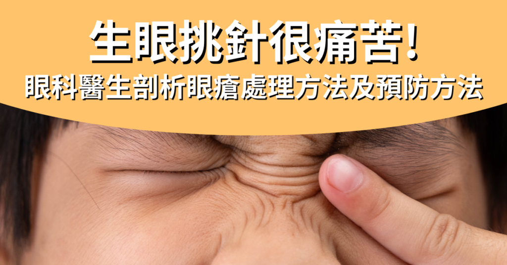
眼瘡 眼挑針 生眼瘡 眼瘡處理 生眼瘡解決方法 眼瘡成因 生眼瘡原因 眼瘡幾耐好 眼挑針初期 內眼瘡 眼瘡自己好 眼生瘡 生眼挑針 眼挑針幾耐好 生眼瘡初期 眼挑針原因 眼倉 眼挑針成因 眼皮生瘡 生眼挑針怎麼辦 眼角生瘡

Single-eye picking is painful! How long will it take to heal? Ophthalmologist discusses treatment methods and prevention methods for eye infections

Does peeking at other people taking a bath cause eye infections or eye picking? How long does it take for the symptoms to improve? Can self-applied ointment cure the condition? Or do you need to see an ophthalmologist to treat the eye infections or eye picking? Many people will undoubtedly have similar questions, especially wondering how long it takes for eye infections or eye picking to improve once they are diagnosed. Before answering these questions, we first need to understand the underlying mechanism of the condition.

Causes of stye/eye pick needle

Eye infections, commonly known as eye picking, are medically referred to as "macula edema." Eye infections/eye picking usually occur near the eyelid glands, causing the eyelids to become red and swollen. The main causes of eye infections/eye picking are excessive gland secretion from the eyelid glands, which leads to blockage of the glands, coupled with a lack of attention to eye hygiene on a daily basis, which leads to bacterial infection.

Additionally, if the patient frequently suffers from eye infections or eye picking, it may also be due to the mites blocking the hair follicles of the eyelashes. These tiny organisms can live in the root of the eyelashes, and when they proliferate in large numbers, they can cause inflammation and the accumulation of pus, ultimately leading to eye infections or eye picking. So it’s not actually caused by peeking into other people’s baths.

For the prevention of eye infections/stye, daily eye care and cleaning are very important, especially for those who frequently wear makeup. It is essential to thoroughly remove makeup and avoid sharing makeup products and tools with others. If eye infections/stye occur frequently almost constantly, consult an ophthalmologist to ensure the condition is properly treated.

Characteristics and symptoms of eye infection/eye picking needle

Stye/stye needle is located in different places and corresponds to different names, namely "external stye" and "internal stye". Stye that is located on the surface of the eyelashes' sebaceous glands is external stye; while internal stye is located within the deep eyelid glands, because the pus is located on the inner side of the eyelid, so the inflammation time is also relatively longer.

In the early stages of the condition, there is only a slight sensation of foreign body in the eye. It does not hurt to touch it. These symptoms will heal on their own with plenty of rest and proper heat therapy. However, if the eye infection is caused by bacteria, the following symptoms may occur:

Symptoms of eye twitching

I. Mild symptoms of eye infection/eye picking needle

  • The eyelids are slightly swollen
  • Mild itching and eye irritation
  • There is a feeling of foreign object.
  • There is a feeling of scratching when blinking.

2. Severe symptoms of eye infection/eye picking needle

  • The eyelids are noticeably red, enlarged, and may even affect vision.
  • Pain, with a slight fever in the eyelid
  • There is a strong sense of foreign object.
  • Short-term visual impairment occurs

If the swelling and pain persists, you should immediately consult an ophthalmologist to treat the eye infection or eye scratch.

How to treat eye bruises/eye pick marks?

Some patients may try to puncture the eye with a needle to burst the eye blister or eye sac, and then squeeze the pus out with a pressure device. However, this practice can lead to bacterial infection, causing the wound to become inflamed again or recur, or causing cellulitis or even brain infection. Therefore, doctors do not recommend this method for patients with eye blisters or eye sac puncture. If you notice that you have eye blisters or eye sac puncture, consult an ophthalmologist. Usually, when treating and managing eye blisters or eye sac puncture, doctors will provide appropriate treatment recommendations based on the severity of the patient’s condition.

I. Mild eye infection/eye picking needle

For minor eye infections or eye picking, if you maintain good eye hygiene and try the warm towel and hot egg method to relieve the pain, the infection or eye picking will subside on its own. Simply boil an egg, peel it, wrap it in a gauze cloth, and gently massage the eyes. Apply this method four to five times daily for 15 minutes each time. This dissolves the blocked oil and pushes it out, reducing the blockage of the oil on the hair follicles. This method will usually resolve the eye infection or eye picking within a few days.

However, if you find that the eye infection/eye pick is too painful, it is recommended that you seek help from a doctor. The doctor will also prescribe some type of steroid or antibiotic based on the condition of the eye infection to help with inflammation and prevent infection.

2. Severe eye infection/eye picking needle

If the eye infection or eyelid tick remains inflamed and does not improve after using the medication, the doctor will recommend that the patient receive injections of corticosteroids to remove the swelling. However, these medications can make the surrounding skin pale, which is not the best option for people with dark skin. Therefore, the doctor may recommend a minor surgical procedure to remove the eye infection or eyelid tick. This type of surgery is relatively simple and minimally invasive, and there is no visible scarring. If the eye infection or eyelid tick continues to occur frequently in the same area, the doctor will send the tissue to a laboratory for cancer screening. However, the eye lid swelling is usually benign and harmless.

How long does it take for eye infections/eye picking to get better?

As for how long it takes for eye infections/eye pickings to improve, it depends on the severity of the patient. Generally speaking, mild eye infections/eye pickings require patience and may take a few days to heal; while severe cases require more patience, with eye infections almost always improving after two weeks. If eye infections/eye pickings are treated with patience, they will almost always improve within a few days, and the course of the disease is also expected to be shortened.

Whether a patient has a mild or severe case, when treating eye infections or eye picking, the most important thing is to seek medical attention promptly and be patient enough to wait a few days to see if the condition improves. It is absolutely not recommended to treat eye infections or eye picking on your own, as this will only lead to more problems and may ultimately require even more patience for the infection to improve and recover. The recovery process may even take over a month.

Can eye infections/eye picking be transmitted?

Generally speaking, eye infections/eye picking are not contagious by themselves. However, if you fail to treat them in time, allowing bacteria or hair follicle mites to grow, this situation may be transmitted to another eye or to another person through contact. Sharing towels, pillows, or cosmetics with patients also increases the risk of infection.

Will eye infections/eye picking needles usually recur if they are not treated?

Everyone who has had a stye or a black eye knows that this sensation is not only extremely uncomfortable but can sometimes directly affect one’s appearance. Although it is not a serious medical emergency, it is definitely a major problem for some people, as styes/black eyes almost always recur. To prevent and prevent recurrence, the most important thing is to start by taking proper hygiene, diet, and preventive measures.

  • Be careful with hygiene: keep your hands clean, especially before touching your eyes; also avoid touching your eyes during the day and wiping your eyes with dirty towels.
  • Healthy lifestyle and diet: Reducing late-night hours to allow the eyes to rest fully can effectively regulate the secretion of oil from the eyelid glands; reducing the intake of fried or spicy foods can reduce the risk of inflammation in the body.

Treating for the prevention of recurrence is more effective for patients with recurrent eye infections/eye picking that are almost resistant to treatment. This type of treatment can kill parasites, is anti-inflammatory and antimicrobial. For example, IPL (strong pulsed light) has light biostimulatory function, effectively stimulating cell mitochondria and promoting cell health. Treatment using tetracycline or tetracycline antibiotics helps to reduce inflammation and kill germs.

If not treated properly, eye infections/stye may recur frequently after a few months, or lead to other more serious eye problems. Therefore, if you have eye infections/stye, the best approach is to seek medical attention from an ophthalmologist to treat them.

Why should I see an ophthalmologist?

Although eye infections are common, doctors can accurately determine the type of infection, assess whether surgery is really necessary, and choose the most suitable incision location and method. Follow-up after surgery is equally important. Doctors provide detailed post-operative care guidance, regularly schedule follow-up visits to assess recovery, and provide preventive recommendations based on your individual situation. This comprehensive treatment and follow-up can truly help you completely resolve your eye infection problem.

💬 Book an eye examination immediately

Persistent eye problems? Book an appointment for detailed assessment and treatment plan.

WhatsApp inquires about eye problems

# #Eye infection treatment: eye drops, #eye pins, eye drops, #eye ointment

Single-eye picking is painful! How long will it take to heal? Ophthalmologist discusses treatment methods and prevention methods for eye infections Read More »

老年黃斑病變會變好嗎?

The ophthalmologist tells you: Age-related macular degeneration is caused by factors and the treatment methods

Macular degenerationAlso known as macular degeneration, this is a chronic eye disease characterized by blurred central vision, distorted images, and dark spots in the center of the field of vision. The macular hole is primarily caused by the gradual aging of the eye as it ages, which is why it is often referred to as age-related macular degeneration.

However, some young people may also be affected by other factors and develop macular degeneration. In the current medical field, is there a feasible treatment option for macular degeneration? Will some macular degeneration cases resolve on their own? Dr. Tong Wenyue explains the causes of macular degeneration and treatment methods: laser photocoagulation, photodynamic therapy, vitreous injection of anti-vascular endothelial growth factor, low vision glasses, bifocal add-on artificial lenses, and more.

What causes macular degeneration?

Age-related macular degeneration occurs due to many factors, the most important of which is age. It is the main cause of visual impairment in the central vision in middle-aged and older adults over 50 years old. However, some young people may also develop macular degeneration; this may be due to congenital genetic factors or adverse habits acquired later in life. Other causes of macular degeneration include:

  • Smoking: Research shows that smokers are 3 to 4 times more likely to develop macular degeneration than the general population.
  • Poor eating habits: Long-term intake of high-fat and high-sugar foods leads to obesity and three types of high blood pressure, affecting the health of the eye’s blood vessels and increasing the risk of macular degeneration.
  • Prolonged exposure to intense sunlight: UVA in the sunlight accelerates the aging of photoreceptor cells, resulting in damage to the macular area.
  • Functions of cell growth factors: Some genes in the human body are abnormal or associated with macular degeneration, such as complement factor B and complement factor H. These abnormal genes have been proven to be highly associated with the development of macular degeneration.

Additionally, although rumors suggest that blue light from electronic devices is one of the reasons for the premature aging of macular degeneration, there are currently insufficient research data to support this theory. However, using electronic devices in darker environments may cause the macular area to absorb more blue light, leading to various eye diseases.

Will the macular degeneration get better?

Some patients ask, "Will the macular degeneration surgery be successful?" or "Will the macular edema resolve on its own?"

It should be noted that macular degeneration cannot be cured on its own; only through certain treatment methods can the condition be slowed down and the symptoms of macular degeneration be alleviated. To prevent vision deterioration, it is recommended to seek the help of an ophthalmologist as soon as possible to treat macular degeneration.

What surgical treatment methods are available for macular degeneration in the elderly?

Treatments for macular degeneration available on the market include laser surgery and injections. The cost/price of both types of macular degeneration surgery depends on the specific treatment plan. Since macular degeneration symptoms may only occur in one eye, but may also appear in both eyes at the same time, the cost/price of treating macular degeneration will vary regardless of whether it is laser or injection surgery.

Currently, intraocular injection is the most common method of treating macular degeneration in older adults. Only doctors who believe that the patient’s condition is severe enough will recommend surgery to treat the macular degeneration.

Additionally, most treatment plans for macular degeneration in older adults focus on more severe wet macular degeneration. As for the treatment of dry macular degeneration, doctors usually recommend that patients eat more foods or supplements that are beneficial for the eyes on a daily basis, avoid smoking and other harmful habits, and undergo regular check-ups.

Laser light coagulation

In addition to treating macular degeneration in older adults, laser photocoagulation can also treat other eye diseases such as cataracts. The principle is to use the heat energy of the laser to eliminate abnormal blood vessel growth in the eye, thereby reducing blood vessel leakage and preventing vision deterioration. However, the photocoagulation treatment for macular degeneration may damage healthy tissue; if the blood vessel is located quite close to the center of the macula, it is recommended to seek other treatment options.

Photodynamic therapy

Photodynamic therapy combines medication and laser to treat age-related macular degeneration. Ophthalmologists first inject a photosensitizing medication into the patient’s arm, which is then transported through the body’s bloodstream and attached to abnormal growing blood vessels. The medication is then activated by cold laser to destroy the blood vessels that are growing in the eye. Unlike laser photocoagulation, photodynamic therapy does not damage surrounding tissues and is also beneficial for improving vision problems. As for the cost of photodynamic treatment for the eyes, feel free to consult our team at any time.

Anti-vascular endothelial growth factor vitreous injection

Injecting medication into the eye is the most commonly used method for treating macular degeneration. Ophthalmologists inject numbing medication into the surface of the eye to prevent abnormal blood vessel growth. This reduces the risk of new blood vessels leaking. The macular degeneration injection treatment not only controls the condition of the macular degeneration but also improves vision damage. The injection treatment for wet macular degeneration is quite effective.

Another method of treating wet macular degeneration with intraocular injections is the injection of steroids. Long-acting steroids can also effectively reduce the damage to the retinal barrier by the blood vessels in the eye and improve macular edema. However, it is important to note that steroids can cause elevated eye pressure and other eye problems such as cataracts.

New technology for treating advanced macular degeneration in the elderly

When laser or injection therapy is not effective in treating macular degeneration, ophthalmologists usually recommend using some auxiliary methods to help improve vision for the patient, such as implanting a bifocal intraocular lens or wearing low vision glasses. Although these methods are more expensive than surgery to treat macular degeneration, they are relatively direct and quick methods.

Low vision glasses

Smart low vision glasses can instantly project images of the surrounding environment onto the retina of low vision individuals in the healthiest area, and adjust according to the patient’s perception. These glasses improve the patient’s perception of the real world by supplementing and correcting visual details. During the setup process, ophthalmologists can intuitively adjust the glasses to help patients find the optimal vision.

Dual-focus add-on artificial crystal

SML (Scharioth Macular Lens) is an implantable lens that can help patients "magnify" their field of vision. It features a specially designed central optical zone that provides high additional optical power of +10.0D, improving near vision without compromising distance vision.

Will some macular lesions heal after surgery?

Unfortunately, macular degeneration cannot be completely cured. Regardless of whether it is performed with macular hole surgery or other treatment methods, it can only slightly improve the vision of the macular hole and prevent the vision from continuing to deteriorate. The key is prevention. During the day, you should take care of your eyes more often, reduce direct sunlight exposure, and supplement with nutrients such as lutein to slow down the aging of the eye. This can reduce the risk of developing macular degeneration.

Diet and care after macular hole surgery

Diet and care after macular hole surgery are crucial steps that directly affect the success rate of the procedure. After macular hole surgery or injection, there are certain precautions to take, such as always following the doctor’s advice to take medication on time and avoid skipping doses. In daily life, it is also necessary to avoid rubbing your eyes, reduce eye strain, and avoid environments with high levels of smoke, such as smoking or construction sites.

In terms of diet, it is recommended to ensure balanced nutrition by eating foods rich in vitamins C, E, A, and zinc, such as dark leafy greens, fruits, and nuts. Typically, doctors will recommend that patients supplement with vitamin C after surgery, as vitamin C can help accelerate recovery and prevent infection; while lutein is recommended as part of long-term care after macular degeneration surgery. Additionally, patients can also try the macular degeneration diet recommended by ophthalmologists or nutritionists to maintain healthy vision.

Dr. Tong Wenjie, an ophthalmologist, reminds patients that if they develop macular degeneration due to high blood pressure or other health issues, they need to make changes to their diet and lifestyle so that their healthy bodies can control the degeneration from worsening and reduce the risk of recurrence.

Common problems

What causes macular degeneration?

Age-related macular degeneration occurs due to many factors, the most important of which is age. It is the primary cause of visual impairment in the central vision in middle-aged and elderly people over 50. Other causes of macular degeneration include smoking, poor dietary habits, and prolonged exposure to intense sunlight.

Is it necessary to stay in the hospital for macular hole surgery?

In most cases, macular hole surgery does not require hospitalization. Generally, macular hole surgery is performed during the day, and patients can go home the same day after the procedure. Regular follow-up visits are scheduled. However, sometimes doctors may decide whether hospitalization is needed based on the patient’s condition.

How many days do I have to stay in the hospital for macular surgery?

Most macular surgery does not require hospitalization; the recovery time after macular surgery can vary from person to person; doctors will provide specific recommendations based on the type of surgery and the patient's condition.

Will the macular degeneration get better?

The effectiveness of macular degeneration treatment varies from person to person, depending on the severity of the condition and the timeliness of treatment. In some cases, improvement can be achieved after treatment, but it will not completely disappear.

Will injections for macular degeneration be effective?

One treatment for macular degeneration is the injection of medication into the macular area of the eye. This treatment can help control the progression of the disease and improve vision, especially in early and intermediate cases. In these cases, injection therapy can have a significant effect.

The ophthalmologist tells you: Age-related macular degeneration is caused by factors and the treatment methods Read More »

老花矯視 presbymax 老花激光矯視 presbymax老花矯視技術 激光矯視 老花 老花激光 老花 激光 激光矯視年齡 激光矯視老花 老花可以做激光嗎 激光老花 激光老花矯視 激光 老花 老花矯視風險 老花手術推薦 老花手術費用香港 激光矯視 改善老花眼的方法 矯視手術 激光矯視 年齡

Get rid of old glasses forever! [Presbymax Laser Cataract Surgery] Helps restore youthful vision

Presbyopia is a vision problem that occurs with age. Almost everyone experiences it, usually in people around 40 years old. Many people with presbyopia are reluctant to wear reading glasses because they fear being ridiculed for being old, which can affect their daily lives. However, nowadays, more and more methods for correcting presbyopia are being introduced to the market. Wearing reading glasses is no longer the only option; for example, Dr. Tong Wenyu, an ophthalmologist who strongly recommends Presbymax laser surgery for presbyopia, not only avoids the embarrassment of wearing reading glasses but also restores the eyes to their youthful vision.

What is presbyopia?

The most common symptom of presbyopia is difficulty reading; text on mobile phones or in books becomes very difficult to see, requiring constant adjustments of distance to find a clearer focus. Prolonged near-vision activities can easily lead to fatigue, dryness, or discomfort in the eyes; working near in darker environments requires more light to help the eyes focus.

The main cause of presbyopia is the degeneration of the lens and ciliary muscles within the eye. This degeneration causes the lens to become rigid and lose elasticity, thereby affecting the eye's refractive power and ultimately impacting vision.

Although cataracts usually occur in people around the age of 40, with the advancement of technology, people are increasingly relying on electronic devices. Prolonged use of screens can cause excessive contraction of the ciliary muscles, leading to the early onset of cataracts. Therefore, protecting eye health is extremely important for modern people. It is recommended to reduce the time spent using electronic devices to give the eyes enough time to relax and slow down the development of cataracts.

2 ways to self-test for farsightedness

Method one: Symptom check

The following are some common symptoms associated with presbyopia; if you have more than 3 of these symptoms, you may have presbyopia.

  • When looking at the phone, you need to hold it farther away to see clearly.
  • When looking at an object, I sometimes want to take off my glasses; wearing them actually makes it clearer to see.
  • You feel that the home or office is dimmer than before.
  • Often misread numbers or letters
  • Often feel tired in the eyes
  • Shoulder and neck pain, stiffness
  • Constantly frowning and feeling dizzy

Method 2: Money banknote test

  1. Take out a banknote and place it in the most visible place for you.
  2. Then focus on the small print on the banknote.
  3. The visual line leaves the banknote and the object 2 to 3 meters away is seen approximately 5 seconds later.
  4. Then look back at the small text line again to see if you can read it in a few seconds.

If you can focus within 1 second, that’s normal; if it takes more than 1 second, you’re at risk of developing presbyopia; if it takes more than 3 seconds, it’s almost certain that you have presbyopia.

If you suspect you have presbyopia, it is recommended to consult an ophthalmologist as soon as possible for professional advice and assistance. The ophthalmologist will determine whether you need to wear reading glasses based on your visual needs, or recommend Presbymax laser surgery to improve your presbyopia. Identifying and treating presbyopia early helps improve vision and enhance quality of life.

What is the difference between farsightedness and presbyopia?

Both farsightedness and nearsightedness cause some difficulty in seeing close objects, but the causes are not the same. Additionally, if you notice vision problems, it is still recommended to have an examination.

  • Presbyopia is a degeneration of the eyeball

Presbyopia is a vision problem related to age. As we age, the lens and ciliary muscles in the eye gradually deteriorate, resulting in a weakened focusing ability that makes it difficult to see clearly near objects and in low-light environments. If presbyopia has severely affected your life, you may consider undergoing Presbymax cataract surgery. This is because Presbymax not only treats presbyopia but also helps improve problems such as nearsightedness, farsightedness, and astigmatism, restoring your eyes to their youthful state.

  • Far-sightedness is innate.

Far-sightedness is a vision problem related to the structure of the eyeball. It is mainly caused by the eyeball being too short or the ciliary muscle being weak. If the eyeball is just too short but the ciliary muscle is strong, there is generally no problem with visual blurring; once the ciliary muscle becomes weak, focusing on near objects requires more effort. Far-sightedness is congenital, meaning most children are farsighted, but there is no need to worry too much; as the eyeball develops normally, the condition will improve.

If you leave your old spectacles lying around, be careful not to cause any health problems!

Many patients find that farsightedness has little impact on their daily lives and are reluctant to address the problem. They believe that they can see clearly with their eyes closed, and even refuse to wear glasses for farsightedness. However, this behavior causes overexertion of the ciliary muscles, exacerbates the strain on the eyes, and leads to conditions such as eye fatigue, discomfort, and pain in the eyeball. In severe cases, it can even spread to the shoulder and neck region, causing symptoms such as shoulder and neck pain, headaches, dizziness, and nausea.

Although cataracts are not a serious condition, the degree of cataracts will increase with age. If cataracts are diagnosed at 40 years old and are 100 degrees, it will increase to 300 degrees by 60 years old. The degree of cataracts will increase by about 10 degrees each year. If the degree of cataracts shows an abnormal decline, this may be a major warning sign of cataracts. Patients should seek an ophthalmologist for detailed examination and appropriate correction as soon as possible. Timely identification and treatment of cataracts is the key to protecting eye health.

What are the methods for correcting farsightedness?

Medical science is constantly advancing, and the methods for correcting farsightedness are also becoming more and more advanced. These include wearing bifocal or multifocal eyeglasses, contact lenses, or surgically implanting artificial lenses, as well as Presbymax laser surgery for farsightedness.

  • Single-focus glasses: Usually, single-focus glasses are only worn when looking at things at close range. These glasses can only provide clear vision within a specific distance. However, if a person also has myopia and hyperopia problems, they need to frequently take off and put on two different-diopter glasses in different situations. In this case, multi-focus glasses can be considered.
  • Multifocal glasses: These glasses are equipped with lenses with multiple focal points, allowing you to see objects at long, intermediate, and near distances with one pair of glasses, eliminating the need to frequently replace your glasses.
  • Multifocal contact lenses: For those who do not like the heavy feeling of wearing glasses or who already have a habit of wearing contact lenses, you can use multifocal contact lenses or have your vision corrected directly through surgery.
  • Surgical treatment: Surgical treatment may include single-eye vision surgery, multifocal intraocular lens surgery, and Presbymax laser correction surgery. Presbymax laser correction surgery is a relatively new option. This procedure reshapes the cornea using laser technology to improve farsighted vision, thereby reducing or eliminating the need for glasses or contact lenses.

Traditional refractive surgery vs Presbymax Presbyopia correction: Which one should you choose?

Single-eye vision surgeryMultifocal artificial lensPresbymax Presbyopia correction
❌ Distorted mid-distance images
❌ Poor sense of three-dimensionality❌ Has a flashing light problem
❌ Not suitable for night drivers
❌ Takes time to adjust✅ Clear vision at long, medium, and short distances
✅ No glare problem
✅ No need for time adjustment; you can resume normal activities within 24 hours after the procedure.

Presbymax cataract surgery mainly involves grinding the cornea with a laser to achieve a multi-focal state. The main operation is to correct the near, far, and presbyopia of the main eye to nearly zero; the other eye retains or creates approximately 150 to 250 degrees of near vision, specifically designed for near vision and to improve presbyopia problems. Additionally, Presbymax incorporates slight spherical aberration to enhance the depth of field in both eyes, lengthen the focal point, and change chromatic aberration, thereby creating a “fusion zone” for both eyes, allowing patients to achieve clear vision at both near and far distances and ensuring the original color and sense of depth remain intact.

Dr. Yu stated that Presbymax is currently one of the most recognized and mature methods for treating presbyopia worldwide. Patients who have undergone Presbymax cataract surgery are extremely satisfied with the results, and the price is also relatively affordable. The surgery takes a very short time, approximately 10 minutes, and patients can resume normal activities within 24 hours. Vision can be fully restored within a few weeks without any discomfort, making Presbymax the best choice for treating presbyopia.

Get rid of old glasses forever! [Presbymax Laser Cataract Surgery] Helps restore youthful vision Read More »

眼有黑點 眼睛有黑點 眼睛有東西在飄 眼球有黑點 眼睛看到固定黑影 眼睛黑點移動 眼睛 黑點 眼睛黑點 眼睛睇野有黑點 眼睛有黑点 眼睛有黑塊 睇野有黑點 眼睇野有黑點 眼有黑影 眼睛看到黑點 眼 黑點 眼睛有黑點怎麼辦 眼睛固定黑點 眼睛有黑點跑來跑去 眼睛見到黑點 眼睛出現黑點

Are dark spots in the eyes a precursor to blindness? Ophthalmologist explains the treatment and improvement methods for fly eye syndrome

Floaters are sometimes very annoying. People who suffer from them see "black spots" floating in their eyes that are neither painful nor itchy, and when you look directly at them they disappear instantly. The shape of floaters varies; for example, they can be black dots, linear, circular, oval, or tadpole-shaped. They are especially easy to spot when looking at bright, clean backgrounds. Floaters are a degenerative eye disease; in mild cases, people generally have no problems with living with the black spots and floating matter in their eyes; daily life is not affected; they do not need to undergo treatment for floaters. However, if the black spots or floating matter in the eyes are too large or too numerous, or if floaters suddenly appear or flash, causing visual impairment, it is necessary to seek ways to improve the situation and consult an ophthalmologist specializing in treating floaters for treatment and surgery or laser treatment for floaters; otherwise, the longer it is delayed, the more serious the problem may become and lead to blindness!

What is fly eye syndrome?

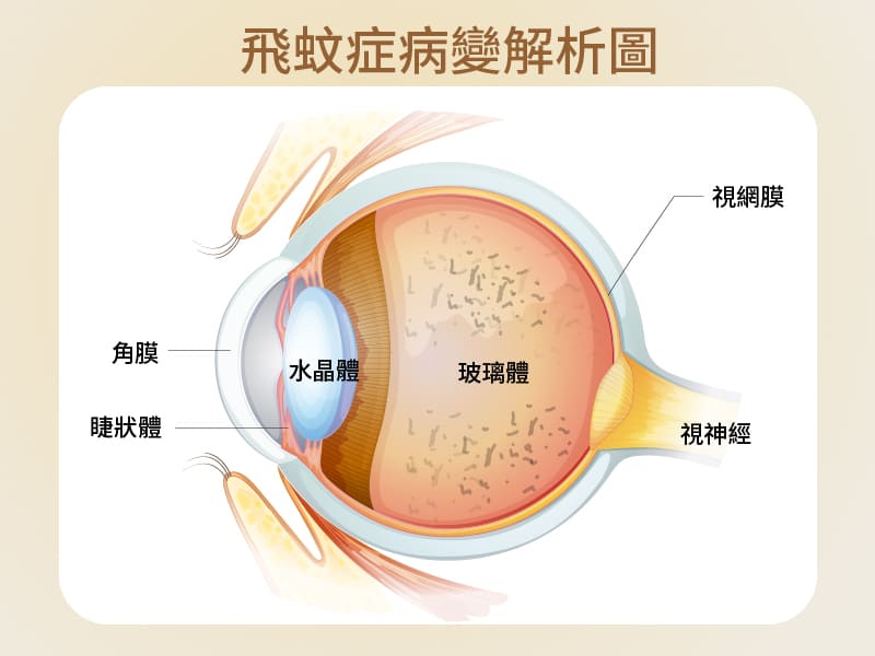
Migraine is medically described as a phenomenon of degeneration of the vitreous humor in the eye. The vitreous humor is a transparent gel-like substance located behind the lens and in front of the retina. Under normal conditions, the vitreous humor fills the entire vitreous cavity to support the shape of the eyeball. However, with age and problems such as nearsightedness, the vitreous humor liquefies and shrinks, forming a cloudy fiber. These fibers then float around in the vitreous humor, and when light rays refract through these fibers, the patient will feel a black spot in the eye. This is a precursor to migraine.

圖解飛蚊症病變

Migraine is medically described as a phenomenon of degeneration of the vitreous humor in the eye. The vitreous humor is a transparent gel-like substance located behind the lens and in front of the retina. Under normal conditions, the vitreous humor fills the entire vitreous cavity to support the shape of the eyeball. However, with age and problems such as nearsightedness, the vitreous humor liquefies and shrinks, forming a cloudy fiber. These fibers then float around in the vitreous humor, and when light rays refract through these fibers, the patient will feel a black spot in the eye. This is a precursor to migraine.

Who is most likely to develop fly-eye?

Floaters usually become more common as people age, so older adults are more likely to experience this symptom of having black spots in their eyes. However, in addition to age factors, other factors that may increase the risk of developing floaters include: nearsightedness, a history of eye surgery, eye trauma, or eye inflammation. Clinical research has found that the high-risk group for floaters includes:

  • Middle-aged and elderly people
  • High myopia
  • Patients with high blood pressure/diabetes
  • Had eyelid surgery
  • Head injuries, such as in car accidents or diving sports
  • Other eye problems, such as: eye inflammation

However, most cases of floaters are benign; factors such as aging and degeneration of the vitreous humor or excessive eye fatigue can all cause the symptoms of floaters. As long as the black spots in the eye do not become more numerous and remain in a fixed position, there is no need to worry too much.

Conversely, if sudden black spots appear in the eyes and affect vision or there is flashing light, it may be vitreous detachment, causing the retina to be pulled into the hole or even detached. This condition must be promptly examined, improved methods sought, or treatment for retinal detachment administered.

Causes of fly eye syndrome

The causes of fly eye syndrome can be roughly divided into three types: physiological, degenerative, and pathological.

  • Rationality

About 80% of cases of floaters are considered to be physiological. Physiological floaters usually occur in people under the age of 40 or those who spend a long time working with their eyes. Most people see black dots in their eyes; these are impurities in the lens that usually do not affect vision and do not require immediate treatment for floaters. Over time, they usually disappear on their own.

  • Degradation

As we age, the vitreous humor in the eye gradually deteriorates, just like the other organs in the body. During this process of degeneration, the vitreous humor forms tiny fibers as it shrinks; these fibers then float in the vitreous humor. When light enters the eye and refracts against these impurities, we see the black spots in our eyes.

  • Pathological

Pathological floaters are caused by eye diseases or systemic vascular diseases. Eye diseases refer to holes (or fissures) appearing in the retina during external forces or as the retina deteriorates, resulting in vitreous hemorrhage and even retinal detachment, which impairs vision. In the most severe cases, it can lead to permanent blindness. Additionally, systemic vascular diseases, such as eye floaters, diabetes, high blood pressure, or macular degeneration, if not treated promptly and aggressively, may also lead to pathological floaters. Patients should seek methods or treatments to improve floaters as soon as possible.

Complications of Glaucoma—Retinal detachment/rupture

Although glaucoma is not a serious disease, retinal detachment caused by glaucoma is an emergency in ophthalmology. One-fourth of patients with glaucoma may suffer from vision impairment due to retinal detachment or retinal tear. Please note that if you experience any of the following symptoms of retinal detachment caused by glaucoma, including:

  • There are a large number of black spots in the eyes within a short period of time.
  • Abnormal flash
  • The dark shadow
  • The line of sight is blocked

This means that the retina may be at risk of tearing, and it is essential to seek medical attention immediately to seek treatment; otherwise, people with fly-eye will suffer permanent damage to their vision or even go blind.

How to treat fly eye syndrome?

If your condition is benign floaters, you do not need to seek immediate treatment. When you notice black spots in your eyes, you can try moving your eyes to allow the fluid in your eyes to flow so that the fibers leave your line of sight. Of course, some people cannot tolerate the floating black spots in their eyes; their attention is often distracted by the black spots, which seriously affects their mood. In this case, you may consider seeking treatment for floaters.

According to current technology, the methods for treating and improving Glaucoma mainly include laser therapy and vitreous removal surgery.

I. Laser treatment for Glaucoma

This treatment method is suitable for larger and concentrated vitreous fibers. It uses laser to shatter these fibers into smaller fragments, improving and eliminating the symptoms of vitreous floaters. The laser treatment is performed under local anesthesia, with no pain during the procedure. The entire treatment takes only 15 to 20 minutes. However, not everyone is suitable for laser treatment to improve vitreous floaters. If the fibers are loose or located too close to the retina or lens, it is not advisable to perform the procedure to avoid complications such as cataracts or damage to the retina.

2. Glaucoma surgery for retinal detachment

Corneal transplant surgery involves removing the cornea from the eye through a small incision and replacing it with a solution to maintain the shape of the eye. The procedure takes about 10 to 15 minutes. However, corneal transplantation cannot always completely remove the fibers; if the procedure itself causes bleeding or retinal tearing, new floaters may form, so most doctors do not recommend using this procedure to treat cataracts.

Methods for improving and maintaining fly eye syndrome

Once fly eye syndrome occurs, it is almost impossible to reverse; even after laser or surgical treatment, there is a possibility of recurrence. What we can do is coexist peacefully with it, or we can improve the symptoms through the following methods to alleviate the condition slightly.

One, daily care for benign floaters

  • Avoid excessive eye strain

Be careful not to strain your eyes too much on a regular basis. Especially for heavy users of 3C devices, you should take a 5-10 minute break every hour to rest your eyes. During this time, blink your eyes and look at the distance to relax your eyes.

  • Good lifestyle habits

In daily life, it is recommended to reduce the time spent on mobile phones; especially, it is important to avoid staying up late at night to use them, ensuring that you get enough sleep and engage in physical activity at the right time are also ways to improve night blindness.

  • Regular check-ups and follow-up

Do not assume that you have benign blepharitis and can relax; patients with blepharitis must undergo regular check-ups to avoid other complications and miss out on the best treatment opportunity.

  • Eye-care eating habits

On weekdays, it is recommended to include a variety of vitamins and foods with antioxidant properties in your diet, such as berries, green or yellow vegetables, carrots, soybeans, milk, and deep-sea fish oil; these are all effective ways to improve night blindness.

2. Postoperative care and maintenance for malignant glaucoma

  • Receiving medical treatment

After the procedure, you must take the medication according to the doctor’s instructions. Remember to thoroughly wash your hands with soap before each dose to avoid cross-infection.

  • Improve your lifestyle

For the first week after the surgery, you should avoid rapidly moving your eyes or overusing your eyes. During this time, try to avoid reading, using a mobile phone, computer, or television. Additionally, it is recommended not to engage in strenuous exercise, climb mountains, dive, or fly on an airplane for 1 month after the surgery, as these activities can stimulate the eyes and affect the speed of post-operative healing.

  • Use eye mask

During the eye repair process, it is recommended to wear an eye mask at all times to prevent collisions and to avoid unintentionally rubbing your eyes during sleep at night. When using it, it is essential to ensure that the eye mask is clean, and it is recommended to wash and disinfect the mask daily to prevent infection caused by bacteria.

  • Daily diet

In addition to avoiding eating irritating foods, it is also important to avoid consuming legumes, as these foods can easily cause nitric oxide in the blood to flow into the eyes, causing the inert gas within the eyes to become bloated and increasing intraocular pressure.

  • Safe home environment

The home should be kept clean and tidy, reduce dust and avoid second-hand smoke. During recovery, do not change the layout of the home or place items on the hallway to avoid collisions or falls that could cause eye irritation in an unfamiliar environment.

  • Regular check-ups

In addition to regular follow-up visits as instructed after the surgery, if you notice any discomfort in your eyes, such as persistent pain, sudden blurred vision, nausea, increased or enlarged shadows, etc., please remember to immediately consult your ophthalmologist to prevent the situation from worsening.

How to prevent fly eye syndrome?

Although we cannot completely avoid the occurrence of retinal edema, we can slow down the aging of the eye and reduce the risk of developing retinal edema through daily care.

One, develop good eye-care habits

  • Avoid excessive use of 3C products (use your eyes for 1 hour, take a 5-10 minute break).
  • Work in conditions of sufficient light and stable lighting without flickering.
  • Avoid staying up late to increase eye strain

Two: Intake of eye-healthy nutrients

  • Carotenoids (deep green, deep yellow and red vegetables)
  • Anthocyanins (Berries)
  • Vitamins (vitamins A, B, C and E)
  • Omega-3 fatty acids (fish oil)

3. Properly control systemic vascular diseases (such as diabetes and hypertension)

  • Maintain a balanced diet and reduce the intake of high-fat, high-salt, and high-sugar foods.
  • Regular exercise (running, cycling)
  • Regularly undergo a comprehensive physical examination

Although most cases of fly eye syndrome do not require treatment, it should not be completely ignored. Regular check-ups are necessary, and if symptoms worsen, seek immediate medical attention to avoid missing the golden period for treating fly eye syndrome.

#Black shadows are floating in and out of my #eyes. The dark spots on my eyes are improving #. There are dark spots in my #eyes.

Are dark spots in the eyes a precursor to blindness? Ophthalmologist explains the treatment and improvement methods for fly eye syndrome Read More »

封面圖_比較_青光眼_黃斑病_白內障

[Treating Eye Diseases] Glaucoma VS Retinal Disease VS Cataract: An Ophthalmologist Explains Eye Diseases Symptoms + Treatment Methods

Elderly people often have blurred vision, which may be due to degenerative problems that prevent them from seeing clearly. But have you ever thought that the problem may be due to eye disease?

Eye disease symptoms

First, it is necessary to distinguish whether the visual acuity is declining suddenly or gradually; the impact of the visual impairment is the intermediate range or the entire image, or is it limited to peripheral vision? Moreover, is there any accompanying pain in the eyes? How can macular degeneration, cataract, and glaucoma be distinguished? In the case of cataract, for example, the visual acuity of older adults generally declines gradually; it also affects the entire image. In contrast, macular degeneration has a sudden, acute decline in visual acuity, affecting central vision. Glaucoma also gradually affects visual acuity; it usually first affects peripheral vision.

These three diseases are easily confused. Older adults can check for them in several ways. One method is using a checkerboard paper. A checkerboard is a square-shaped paper that older adults can use to check whether the lines have changed into curves. This can help determine whether macular degeneration is present. For other problems, an eye specialist’s examination is required to determine the exact cause of the eye condition.

How to cure it?

Diabetic retinopathy generally has two complications: hypoxia, which leads to the growth of blood vessels; and leakage of those fragile blood vessels, which can lead to vitreous hemorrhage or macular edema. To prevent this, injections of eye medications are usually administered to stop the growth of blood vessels and thereby reduce hypoxia.

Glaucoma is basically treated with minimally invasive cataract surgery to remove the cloudy lens and implant an artificial lens. Lastly, glaucoma is controlled through early treatment by controlling the nerve fibers and preventing them from continuing to damage the optic nerve. There is no treatment for glaucoma; only through testing, medication, surgery, or laser to control eye pressure can slow the damage to the optic nerve.

Older adults can prevent eye diseases by changing their daily habits, such as eating less sugar and salt; eating foods rich in vitamins A, carotenoids, or omega-3 fatty acids, which have antioxidant effects; doing at least 30 minutes of aerobic exercise; and not smoking. Older adults should have regular eye exams to prevent and treat eye diseases early. Avoiding exposure to ultraviolet light as much as possible can prevent various eye diseases.

Source of data:Topic

[Treating Eye Diseases] Glaucoma VS Retinal Disease VS Cataract: An Ophthalmologist Explains Eye Diseases Symptoms + Treatment Methods Read More »

封面圖_眼睛紅痛_隱疾_風濕關節炎_眼發炎

[Inflammation of the eyes] Red, painful eyes or other signs of illness: doctor: 80% of patients with rheumatoid arthritis have inflammation of the eyes

Many doctors use the eyes as a window into the body's health condition. Especially for long-term patients, which diseases can affect the eyes?

Diseases that affect the eyes

Conditions such as eye pain, red eyes, and inflammation of the eyes are generally experienced by older adults. However, if the condition persists, we may worry that there is another underlying health issue. One of these is an autoimmune disease, which causes various inflammatory conditions in the body. The eyes are also one of the affected organs; if left untreated or not addressed properly, they can lead to continued visual impairment and even the risk of blindness.

Symptoms and treatment of immune system diseases

Immune system diseases occur in people of different ages. The most common are rheumatoid arthritis, dry joint syndrome, ankylosing spondylitis, or lupus erythematosus. In the case of rheumatoid arthritis, patients may experience pain in the lower back, hip pain, or a feeling of tightness in the back when getting up due to inflammatory symptoms. Due to inflammation, calcification occurs. The joints become fused together, making it difficult to get up in the morning or to reach your toes when bending down; this can affect joint function over time. The first line of treatment is systemic therapy, usually prescribed by a rheumatologist. Oral anti-inflammatory medications are usually prescribed.

In first-line treatment, steroids are commonly used, but if we use high doses of steroids or continuous treatment, we generally switch to second-line drugs to reduce the side effects caused by steroids. In this case, biopharmaceuticals and immunosuppressants are used as second-line drugs to control long-term treatment.

Research shows that 80% of patients have at least one episode of eye inflammation in their lifetime. Conjunctivitis is one of these inflammations, which releases cells that cause visual impairment. These cells combine with proteins in the eye and accumulate in the anterior chamber, causing the anterior chamber fluid to not drain away, leading to increased eye pressure, damage to the optic nerve, and the development of glaucoma.

To treat the inflammation, a blood test is usually performed to look for inflammatory factors and X-rays are taken, such as X-rays of the lungs or the lumbar spine, to see if there are any problems with the lungs or the lumbar spine. If so, medication is usually used to control the inflammation (a type of anti-inflammatory steroid eye drop). Additionally, to check for any side effects or complications, eye drops for glaucoma are used to control the eye pressure, so ophthalmologists and rheumatologists work together to care for and treat the patient.

Remedy

There are different types of eye drops; some are ordinary moisturizing eye drops that do not help the condition; sometimes they can even delay treatment; others have the effect of constricting blood vessels, but they do not control inflammation, so it is possible to mistakenly believe that the condition is continuing to improve; finally, if the treatment dose is insufficient or the time of administration is insufficient, it will delay treatment, causing irreversible problems in the eyes.

Coronavirus

Sinusitis is accompanied by respiratory infections such as the flu or a viral cold. In fact, it is also one of the symptoms of COVID-19. The most common symptom of sinusitis is the swelling of the conjunctiva around the eye, or a condition of redness and inflammation. Research shows that 30 COVID-19 patients underwent an examination during their hospitalization, and one of them was found to have symptoms of sinusitis, so there is a close connection.

To prevent COVID-19, first of all, we must wash our hands frequently, using water and soap for at least 20 seconds. Additionally, we need to maintain appropriate social distancing of 1.5 meters, and we need to maintain good air circulation to protect ourselves from COVID-19.

Source of data:Topic

[Inflammation of the eyes] Red, painful eyes or other signs of illness: doctor: 80% of patients with rheumatoid arthritis have inflammation of the eyes Read More »

封面圖_新冠病毒病徵_紅眼症_湯文傑眼科醫生

Is red-eye syndrome also a symptom of the novel coronavirus disease?

Is red-eye syndrome also a symptom of the novel coronavirus disease?

With the COVID-19 pandemic raging, people are focusing more on prevention, infection symptoms, and rehabilitation treatment, and less on the relationship between infection and visual risk. In fact, red eye syndrome is also an early sign of the novel coronavirus, and people are concerned about whether droplets can be infected by falling into the eyes, and whether wearing glasses has any effect on prevention. The following is a detailed answer from an ophthalmologist.

In mild cases of COVID-19, common symptoms include runny nose, headache, fatigue, sore throat, cough, and even muscle aches and vomiting. It is also believed that red eye syndrome (the medical term is conjunctivitis), which is an early sign of the novel coronavirus, is also a common symptom of COVID-19. Which is true or false? OphthalmologistYu WenjieExplanation: Red eye syndrome or conjunctivitis is accompanied by upper respiratory tract infections (such as colds and flu). It may also be a sign of infection with the novel coronavirus. The common symptoms of conjunctivitis are the symptoms of swollen or red eyes and inflammation; a study published in the Journal of Medical Virology examined 30 patients admitted for COVID-19 and found that one patient had conjunctivitis.

Red-eye syndrome can cause various eye diseases.

According to the aforementioned study, although the incidence of conjunctivitis in patients is lower, in addition to conjunctivitis, reports indicate that the novel coronavirus is associated with other eye problems, including external epithelial membrane inflammation, choroidal inflammation, inflammation of the lacrimal glands, inflammation of the retina and optic nerve, although these are also rare, some of these problems can lead to loss of vision.

Residents wear masks when going out in their daily lives, but they are most concerned about the possibility of the novel coronavirus virus spreading to the eyes, causing infection. Dr. Tu said that the transmission of the virus to the eyes has not yet been thoroughly studied; however, the mucous membranes in many body cavities and organs, including the respiratory tract, are usually the most susceptible to infection with the novel coronavirus and viruses. The surface of the eyes and the inner eyelids are also known as the mucous membranes of the conjunctiva, so if the infected droplets fall into the eyes, they may easily become infected. Published reports indicate that the novel coronavirus 2 (SARS-CoV-2) may be transmitted through aerosol contact with the mucous membranes.

Eye protection goggles are a valuable asset

It was thought that glasses and sunglasses could act as a barrier, but it was actually just a “misunderstanding.” Dr. Tu said, “Both glasses and sunglasses cannot completely block respiratory droplets sprayed toward you; instead, protective goggles on the sides and around the eyes provide better protection.” In fact, the transmission method of the novel coronavirus is still believed to be primarily through respiratory droplets transmitted between people, but the virus can survive on surfaces for several days. Therefore, touching infected surfaces and then touching the eyes, nose, or mouth without washing hands can lead to infection, but this risk is generally considered very low.

Cleaning the eyes with water or over-the-counter eye drops can reduce the risk of infection. He points out that the eyes have their own protective mechanisms, including antimicrobial proteins and natural lubrication. Therefore, daily cleaning or rinsing the eyes with water is unnecessary and may actually wash away some of the natural protective barriers on the eye surface, thereby increasing the risk of infection. If the eyes become inflamed or red, using over-the-counter artificial tears may help alleviate the symptoms; however, if the symptoms persist, it is recommended to seek medical attention as soon as possible.

Remove contact lenses to maintain hygiene

Not only have people with myopia said that wearing and removing contact lenses frequently increases the risk of contracting the novel coronavirus, but Dr. Tu has given reassuring words. Under appropriate sanitary conditions, there is no evidence that wearing contact lenses increases the risk of contracting the novel coronavirus. However, contact lens wearers touch their eyes more frequently than the general population. The American Academy of Ophthalmology recommends that contact lens wearers temporarily switch to wearing glasses to reduce the risk of contracting the novel coronavirus. Additionally, glasses can partially reduce the risk of irritation caused by wearing contact lenses and serve as a barrier that prevents touching the eyes before stopping. Although not the best protection, glasses can also serve as a partial protective cover against respiratory droplets.

Despite this, the American Optometric Association states that as long as proper daily care is taken when wearing contact lenses, ensuring hygiene and washing hands frequently, the problem can be resolved. If the eyes become red, the wearing of contact lenses should be stopped and medical attention should be sought.

5 Major Ways to Prevent COVID-19 Infection

To reduce the risk of contracting the new coronavirus, Dr. Yu has the following recommendations:

  1. Clean your hands frequently
    - Wash your hands with soap and water for at least 20 seconds.
    - Try to avoid touching your eyes, nose, and mouth as much as possible.

  2. Reduce contact
    - Maintain social distance; please use the length of two arms (about 6 feet) as the guiding distance.
    - Try to avoid entering indoor or outdoor spaces with poor air circulation.
    - If you are ill, please stay at home unless you need medical care.

  3. Covering coughs and sneezes
    - When coughing or sneezing, please use a tissue to cover your mouth and nose.
    - Immediately wash your hands with soap and water for at least 20 seconds after coughing or sneezing.
    - Wearing a mask in public places to cover the mouth and nose.

  4. Clean and disinfect the surface of objects
    - Current evidence indicates that the novel coronavirus can survive on the surfaces of various materials for several hours or even days. Cleaning visibly dirty surfaces and then disinfecting them in a home or community setting is the best way to prevent the novel coronavirus and other viral respiratory diseases.

  5. Getting vaccinated with the COVID-19 vaccine
    -Vaccination has been shown to significantly reduce the risk of infection with the novel coronavirus and alleviate the severity of symptoms.

Is red-eye syndrome also a symptom of the novel coronavirus disease? Read More »

Visual and visual migraine: what is the difference?

Occipital migraine is a rare disease characterized by temporary loss of vision in one eye, or even temporary blindness. Occipital migraine is caused by reduced blood flow to the retina or the blood vessels behind the eye.

In ocular migraine, the patient’s visual acuity usually returns to normal within an hour. Ocular migraine can be painless or occur simultaneously with (or following) migraine.

Unfortunately, the term “ocular migraine” is often used to describe a more common (harmless) condition—called visual migraine or migraine premonition—whose characteristic is temporary visual impairment that usually disappears within 30 minutes.

Unlike ocular migraine, visual migraine usually affects both eyes.

Now let's take a closer look at ocular migraine and visual migraine:

Symptoms of ocular and visual migraine:

Symptoms of ocular migraine typically include a small blind spot that affects the central vision of one eye. This blind spot becomes larger, making it unsafe to drive or to read with the affected eye.

In some cases, the entire field of vision in one eye may be affected. Usually, this condition lasts for less than an hour.

The visual migraine symptoms may vary, and may include:

1. Flashing blind spots near the center of the field of view or near the center

2. Colored light waves around the central blind spot in the form of a wave or a serrated pattern

3. Blind spots that move slowly across the entire field of view

Symptoms of visual migraine usually affect both eyes and last for 30 minutes or less. Soon after the visual migraine symptoms subside, a headache may occur, or there may be no headache at all.

If you experience a blind spot or other visual impairment and are unsure whether it is ocular migraine or visual migraine, please cover one eye at a time. If only one eye experiences visual impairment, it is most likely ocular migraine. If it affects both eyes, it may be visual migraine.

However, do not take any risks. If you suddenly encounter any blind spots in your field of vision, please immediately call or consult an ophthalmologist to determine whether it is harmless or a sign of a more serious condition (such as retinal detachment).

What causes eye and visual migraine?

People believe that ocular migraine and migraine are caused by the same underlying causes.

Migraine has a hereditary basis; some studies show that up to 70% of people with the disease have a family history of the disorder.

According to the World Health Organization, migraine appears to be caused by activation of deep brain mechanisms that release inflammatory substances around the nerves and blood vessels in the head and brain.

Imaging studies have found that during migraine headaches and pre-migraine headaches, blood flow to the brain changes. However, the reason for this phenomenon and the causes of spontaneous resolution of migraine headaches and visual migraine headaches remain unknown.

Common trigger factors for migraine attacks that may lead to a person experiencing a migraine include certain foods, such as aged cheese, caffeinated beverages, red wine, smoked meat, and chocolate.

Food additives such as monosodium glutamate (MSG) and artificial sweeteners can also cause migraines in some people.

Other potential triggers of migraine include cigarette smoke, perfumes and other strong odors, bright lights or flashing lights, lack of sleep, and emotional stress.

Treatment and prevention

As already noted, visual impairments caused by ocular migraine and visual aura usually disappear within an hour or less without requiring treatment.

If the task you are performing requires clear vision, if you experience eye or visual migraine, stop what you are doing and relax until your vision returns to normal.

If you are driving, please stop on the side of the road and wait until the visual impairment is completely resolved.

If you experience visual impairment accompanied by headaches, consult your family doctor or neurologist to evaluate the onset of your headaches.

Your doctor can provide you with advice on the latest medications for treating migraine headaches, including medications designed to prevent future attacks.

It is best to record your diet and activities before the onset of ocular or pre-ocular headaches to see if you can identify any triggers that might be avoided in the future.

If your migraine or pre-migraine (visual migraine) seems related to stress, you can reduce the frequency of your migraines without medication by following these simple methods:

. Regularly eat healthy meals

. Avoid common causes of migraine headaches

. Adequate sleep

. Try methods to relieve stress, such as yoga and massage.

Visual and visual migraine: what is the difference? Read More »

My eyes suddenly turned red. That's what I should do.

Red eye indicates some form of inflammation of the eyes. This condition is usually benign and self-limiting. Recognizing the warning signs of urgent referral to an ophthalmologist is crucial to prevent long-term complications. When patients experience severe eye pain, copious purulent secretions, loss of vision, distorted pupils, severe photophobia, external eye injury, and herpes infection, referral is necessary. The cause of red eye can be diagnosed through a detailed medical history and a careful eye examination.

Bleeding or swelling
Is there any pain?
Has the vision been affected?
Are there any other complications?

Treatment options for red eye in certain cases:

1. Viral conjunctivitis: Treatment is supportive and may include cold compresses and artificial tears. Local antibiotics are rarely used because secondary bacterial infections are uncommon. One study showed that SARS-CoV-2 can be transmitted through droplets, but infected patients are unlikely to transmit the virus through tears. Researchers from the National Infectious Diseases Centre in Singapore collected tear samples from 17 COVID-19 patients from the initial symptoms until recovery, which usually occurred on the 20th day. All patients showed no eye symptoms, but one patient developed conjunctival swelling during hospitalization. No virus was detected in the 64 tear samples collected during the study, however, viral load was detected in nasal and throat swabs.

2. Bacterial conjunctivitis:Antibiotics are the mainstay of treatment; the choice of antibiotics should be based on cost-effectiveness and local patterns of bacterial resistance.

3. Chlamydia conjunctivitis:Treatment methods include topical use of erythromycin eye ointment, as well as oral treatment with azithromycin (a single 1-gram dose) or potentomycin (two times daily, 100 mg each time, for 14 consecutive days) to clear up genital infections; the patient's sexual partner also needs to be monitored and treated.

4. Allergic conjunctivitis:Avoiding contact with allergens and using artificial tears are effective ways to relieve the symptoms of mild cases. Non-prescription antihistamines/vasoconstrictors are also viable options, sometimes requiring the use of topical steroids to treat acute attacks.

5. Dry eye syndrome:Treatment includes frequent application of ointment during the day and night, as well as the insertion of artificial tears to reduce evaporation rates. New treatment methods include topical ocular drops containing phospholipids that increase tear production. Omega-3 fatty acids have also been shown to be helpful.

6. Conjunctivitis: There has not yet been shown to be a significant benefit of topical NSAIDs compared to placebo in the treatment of conjunctivitis; topical corticosteroids may be useful in severe cases; however, they are usually self-limiting.

7. Subconjunctival hemorrhage: We can use only heat packs and ophthalmological lubricants; recurrent bleeding may require a physical examination.

My eyes suddenly turned red. That's what I should do. Read More »