Causes, Symptoms, and Treatment Methods of Glaucoma

Glaucoma is a chronic eye disease that has been dubbed the "sight thief." There are no early warning signs; early glaucoma has no obvious pain or symptoms, making it difficult to detect. It is only noticed when the patient's vision has narrowed. However, when the patient notices that their vision is narrowing, their eyes have already suffered irreversible damage. According to the Hong Kong Ophthalmological Society, 23% patients who lost their sight were blind due to glaucoma; those with farsightedness and those over 40 years of age are at high risk of acute glaucoma.

Cataract occurs when the pressure inside the eye becomes too high, causing gradual damage to the retina, the optic nerve, and the optic fibers. However, some patients have eye pressure within normal ranges (10-21 mmHg), but the optic nerve is unable to withstand the pressure and is damaged, resulting in a narrowing of vision. Early loss of vision usually begins in the peripheral vision and eventually only remains in the central portion. If no appropriate treatment is provided, vision will gradually decline or even be completely lost.

Deepen our understanding of the causes behind Glaucoma

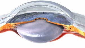
In a healthy state, our eyes continuously produce a liquid called “aqueous humor.”Maintain a healthy eye pressure,The aqueous humor must be drained from the eyes. The cause of cataract is excessive eye pressure, which gradually damages the optic nerve. However, some patients have normal eye pressure (10-21 mmHg), but the optic nerve still cannot withstand the pressure and is damaged, resulting in blurred vision. Early vision loss usually begins in the peripheral vision and eventually only remains in the central part. If proper treatment is not provided, vision will gradually decline or even be completely lost.

Ophthalmologist Tong Wenyue Explains the Symptoms of Glaucoma

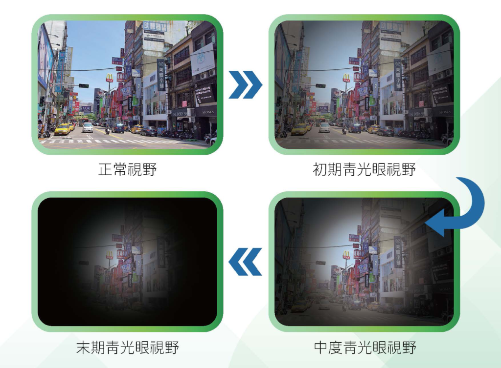
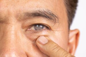
Illustration of Glaucoma Symptoms

Types of Glaucoma

According to its characteristics and causes, cataract can be divided into different types. These include chronic cataract and acute cataract.

  • Chronic Glaucoma
    • In the early stages, there are no obvious symptoms; the patient may not notice any signs. As the disease progresses, the patient’s field of vision gradually shrinks, eventually leaving only central vision; peripheral vision is lost, resulting in what is known as “tunnel vision.” If the condition continues to worsen, it can lead to complete blindness.
  • Acute Glaucoma
    • Sudden increases in eye pressure can cause noticeable symptoms such as blurred vision, seeing rainbow-colored rings when looking at light, red eyes, blurred cornea, severe eye pain, headaches, and nausea and vomiting. If acute glaucoma is not treated promptly, it can even lead to permanent vision loss within a day.

Depending on the cause, glaucoma can be divided into primary and secondary types.

  • Primary glaucoma
    • No obvious causes are identified, including open-angle glaucoma and angle-closure glaucoma.
      • Angle-closure glaucoma refers to the opening of the anterior chamber, but elevated eye pressure gradually damages the optic nerve. In the early stages, most patients fail to notice any symptoms or signs.
      • The characteristic feature of an angle-closure glaucoma is the abnormal closure of the anterior chamber, which causes the outflow of aqueous humor to be obstructed and the eye pressure to rise. If the anterior chamber closure is slower, the rise in eye pressure is slower, and the patient may not experience any symptoms.
  • Secondary glaucoma
    • Elevated eye pressure caused by other diseases, such as mature cataracts, conjunctivitis, intraocular hemorrhage, tumors, trauma, post-surgical complications, and improper use of corticosteroids. There are many types of glaucoma, but early detection and timely treatment are key to reducing the risk of vision damage.

Symptoms of different types of glaucoma

Want to know if you have Glaucoma? In addition to being able toComparison of symptoms of different types of eye diseasesIn addition, we can also gain a deeper understanding of the symptoms of different types of glaucoma.

Early chronic open-angle glaucoma often has no obvious symptoms, but as the disease progresses, patients may gradually experience loss of peripheral vision.

Early symptoms of acute angle-closure glaucoma may include eye pain, eye redness, blurred vision, eye swelling, and nausea and vomiting. Attacks of acute angle-closure glaucoma can lead to acute severe eye pain, headache, nausea and vomiting, visual blurring, and the appearance of halos.

Causes and risk factors of Glaucoma

Age 40 or older

Older age is an important risk factor for cataracts. As age increases, the incidence of cataracts increases exponentially. People over the age of 60 are six times more likely to develop cataracts.

High blood pressure

People with high blood pressure are twice as likely to develop “normal-pressure glaucoma” than others. High blood pressure damages the eye’s blood circulation system, causing abnormal blood flow in the eye. Therefore, even if the eye pressure is within normal limits, high blood pressure still accelerates the progression of optic nerve degeneration.

Diabetes

The results of the study showed that compared with non-diabetic patients, people with diabetes were twice as likely to develop glaucoma (mainly open-angle glaucoma).

Having cardiovascular diseases

People with cardiovascular diseases are at a high risk of developing glaucoma. Although high blood lipid levels do not directly cause eye disease, research shows that high blood lipid levels are associated with increased eye pressure, which in turn increases the risk of developing glaucoma.

Previously used for taking or applying steroids externally

Long-term use of steroids can lead to elevated eye pressure, which in turn can cause steroid-induced glaucoma. Prolonged use of steroids blocks the drainage channels in the eye, reducing the rate of drainage of the aqueous humor, which in turn causes elevated eye pressure. However, once steroids are discontinued, the eye pressure in most patients can return to normal levels.

Histology of the Glaucoma Family

Having a family history of glaucoma carries a higher risk. Aperture-type glaucoma is the most common hereditary form of the disease. If a close relative has glaucoma, your risk of developing the disease will increase by 4 to 9 times.

Eye injury

When the eye is hit by a hard object (especially blunt trauma), it may cause the eye structure to hemorrhage, block the flow of aqueous humor, and lead to increased eye pressure, which in turn can cause glaucoma.

Chronic migraine

Long-term migraine headaches are closely related to glaucoma. Glaucoma is generally divided into two forms: acute and chronic. The majority of cases of migraine are caused by chronic glaucoma. On the other hand, migraine is sometimes misdiagnosed with acute angle-closure glaucoma. This is because the outflow channel of the anterior chamber of the eye becomes blocked for a short period of time, causing a rapid increase in eye pressure, accompanied by corneal edema, which can lead to severe headache symptoms. This condition is often easily mistaken for migraine and delays timely treatment of glaucoma.

Far/near vision

Both nearsightedness and farsightedness are among the causes of glaucoma. In addition to angle-closure glaucoma, patients with high farsightedness are more likely to develop this condition. Research also indicates that nearsighted eyes are more susceptible to increased eye pressure. Especially those with a near-sightedness of more than 500 diopters and who also experience increased eye pressure are more likely to experience a narrowing of the visual field.

Sleep apnea

Recent literature studies have indicated a relationship between sleep apnea and glaucoma. It is possible that patients experience a lack of oxygen in their bodies during sleep, which in turn affects blood circulation around the eye, leading to oxygen deficiency in the optic nerve, resulting in gradual visual impairment.

Glaucoma examination

Why should I have an eye exam regularly?

Glaucoma is a common eye disease that gradually damages the optic nerve, eventually leading to vision loss. However, early chronic glaucoma often has no obvious symptoms. Many patients only realize they are affected when the disease has progressed to a late stage. Once the optic nerve is damaged, it cannot be reversed. Therefore, regular glaucoma screenings are crucial for early detection and control of glaucoma. Not only can they help in early diagnosis and treatment to prevent vision loss.

Suitable for
  • Age 18 and above (especially 40 years or older)
  • Deep myopia (600 degrees or above)
  • People with diabetes
  • The family has a history of glaucoma.
  • The eye has been injured
  • Suspected of having Glaucoma
Comprehensive eye examination scope
1. Examination and inquiry of medical records

Understand the signs of visual problems and eye diseases

2. Visual acuity and refraction testing

Through various procedures and instruments, the condition of the patient's vision can be accurately assessed, including nearsightedness, farsightedness, astigmatism, and presbyopia.

3. Eye pressure measurement

Eye pressure is one of the main indicators of glaucoma. Doctors usually use non-contact pressure gauges or instruments that apply slight pressure to measure eye pressure. This process is usually painless and can be performed in an ophthalmology clinic.

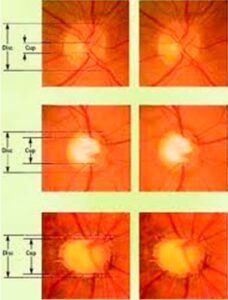
4. Visual nerve examination

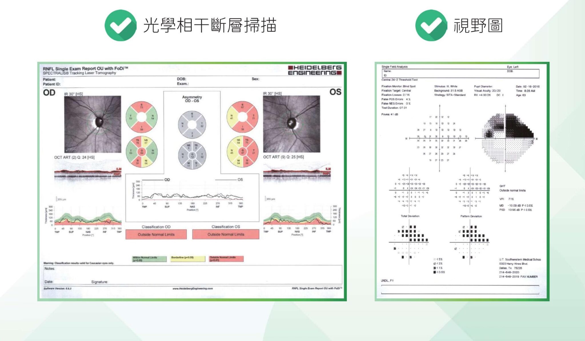
In the diagnosis of glaucoma, optical coherence tomography (OCT) plays an important role. This is an non-invasive optical diagnostic technique that doctors use to examine the eyes to determine the presence of signs of optic nerve damage. Through advanced equipment, it is possible to more accurately assess the health of the optic nerve and provide a more comprehensive assessment to detect glaucoma early. By detecting visual nerve damage at an early stage and developing appropriate treatment plans.

Glaucoma examination
 5. Visual field examination

Visual field testing can help doctors assess the integrity of the visual field and any potential visual defects. This test is usually performed using an automatic visual field tester, where the patient is asked to focus on a specific point and detect changes in the brightness of the light spots in all directions. This can help the doctor determine if the visual field is damaged.

6. Mirror inspection

Eye examinations include using lenses to assess the structure of the front and back of the eye. Doctors can use eye examinations to check the health of areas such as the iris, the lens, and the retina. This examination can provide important information about the internal structure of the eye.

7. Analysis and construction

Treatment methods for Glaucoma

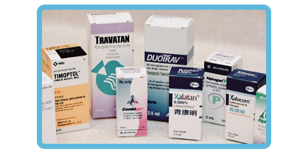
Prescription eye drops are one of the common methods for treating glaucoma. They can be used to control eye pressure levels to an appropriate target range, protect visual nerve tissue, and reduce further visual damage. As a non-invasive treatment method, the side effects from eye drops are relatively low, and they are also safer than laser and surgical treatments. Currently, many glaucoma eye drops are available in combination formulations, allowing patients to use only one drop of eye drops per day.

Currently, many Glaucoma medications are available in combination formulas, so patients only need one drop of eye medication per day.

If the condition of ocular hypertension cannot be controlled with drug treatment, patients can consider laser and surgical treatment, such as: Xia Liang resection, drainage surgery, conduit implantation, conjunctival photocoagulation or cryotherapy, argon laser periorbital shaping, and micro-invasive surgery for ocular hypertension.

  • The Xia Liang resection is a surgical method that removes part of the small-liang tissue to improve the drainage of the aqueous humor and thereby reduce intraocular pressure.
  • Drainage surgery involves creating a new drainage channel in the eye to facilitate the drainage of the aqueous humor.
  • Endovascular implantation involves inserting an outflow conduit into the eye to guide the fluid out of the eye.
  • Mesothelioma photocoagulation or cryotherapy is a procedure that uses laser or freezing technology to damage the mesothelial tissue, reducing the production of aqueous humor and thereby lowering intraocular pressure.
  • Argon laser perirhinal reshaping is a procedure that uses argon laser to expand the aqueous outflow channel of the eye, increasing the amount of aqueous outflow.

New options for patients with mild to moderate glaucoma: the latest treatment methods for glaucoma

Micro-incision Glaucoma Surgery (MIGS)

In recent years, micro-incision glaucoma surgery (MIGS) has begun to be used, which is less invasive than traditional glaucoma surgery. This reduces the potential for complications and provides a new option for patients with mild to moderate glaucoma. MIGS has higher safety and faster recovery times, while reducing the burden on patients with glaucoma medications.

Selective laser fine-beam shaping (SLT)

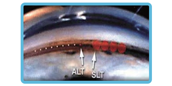
Selective laser trabeculoplasty (SLT) is a clinic procedure that can reduce intraocular pressure in patients with glaucoma. The laser is directed through a special lens into the outflow system of the eye, stimulating biochemical changes there, thereby improving the outflow of aqueous humor from the eye. Because SLT has excellent “benefit-risk” characteristics, it has been an option for the treatment of glaucoma early in the treatment strategy (including as the primary treatment).

Studies comparing SLT and eye drops as the primary treatment found that the treatment effects between the two groups were similar. Although some patients still needed eye drops after SLT, they needed fewer eye drops to control their glaucoma.

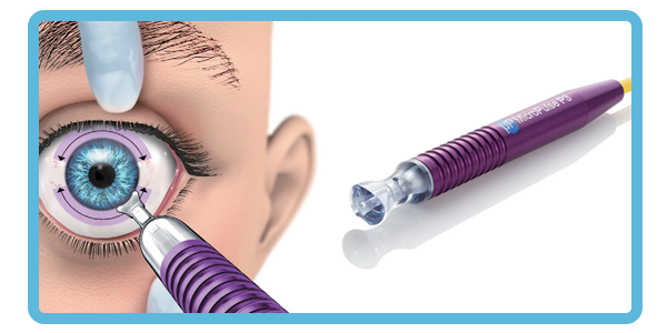
MicroPulse®P3

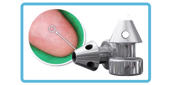
The MicroPulse® P3 Glaucoma Device (MP3) is treated by the CYCLO G6 Glaucoma Laser System. Using non-pulsed, repeatable and predictable MicroPulse technology, it provides innovative photocoagulation that destroys the corneal tissue, reduces aqueous humor production and thereby lowers intraocular pressure.

Common Questions About Glaucoma

What can you eat to prevent cataracts?

It is recommended to increase the intake of vegetables and fruits rich in vitamins, minerals, and antioxidants, such as kale, carrots, and peaches. Additionally, eating foods rich in nitrogen, such as deep-green vegetables, can also help reduce the risk of cataracts.

How long after being diagnosed with Glaucoma will I go blind?

Glaucoma does not necessarily lead to blindness; if the patient receives stable treatment, the condition can be effectively controlled. The probability of blindness caused by glaucoma is between 10% and 25 percent. In the case of acute glaucoma, if treatment is not provided in time, patients with acute glaucoma may face the risk of blindness within a few days.

Chronic glaucoma usually has no obvious symptoms in its early stages. When problems such as blurred vision, being unable to see objects clearly at a certain angle, and tripping over stairs occur, it usually indicates that the disease has entered its late stage, and the patient may face the risk of blindness within 3 to 5 years.

Can Glaucoma be cured?

There is currently no cure for keratoconus, but by performing proper diagnosis and treatment at an early stage and working closely with an ophthalmologist, it is possible to maintain good eye health to the greatest extent possible.

How to detect glaucoma early?

Early glaucoma lacks obvious symptoms; once a patient notices that their field of vision is narrowing, the eye has already suffered irreversible damage. Therefore, adults are advised to undergo comprehensive eye examinations every one to two years to check for eye pressure, the cornea, and other eye health conditions, to avoid missing the opportunity to control the disease.

Consultation registration