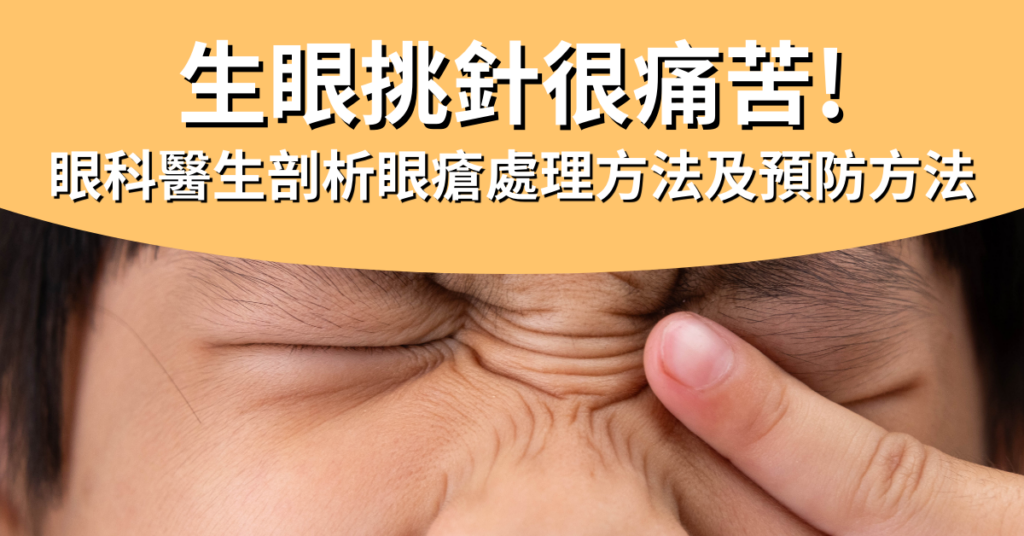
Peeping at others while they bathe can cause styes or chalazia? How long do the symptoms last? Can they heal with self-applied ointment, or should one see an ophthalmologist for treatment? Many likely share these questions, especially the burning desire to know: once afflicted, how long will a stye or chalazion take to resolve? Before addressing these concerns, we must first understand the underlying mechanism.
Causes of Styes/Stye Formation
Stye, commonly known as a "pimple on the eyelid," is medically termed a "hordeolum." Styes typically develop near the meibomian glands and can cause eyelid redness and swelling. The primary cause of styes is excessive oil secretion from the meibomian glands, leading to gland blockage. This is often compounded by poor eye hygiene practices that allow bacterial infection to occur.
Additionally, if a patient frequently develops styes or chalazia, it may be due to eyelash follicles being blocked by mites. These tiny organisms can live at the base of eyelashes, and when they multiply excessively, they may cause inflammation and pus accumulation, ultimately forming styes or chalazia. So it's actually not caused by peeping on others while they bathe.
To prevent styes, daily eye care and hygiene are crucial, especially for those who frequently wear makeup. Thoroughly remove all makeup and avoid sharing cosmetics or makeup tools with others whenever possible. If styes recur frequently and persist for extended periods, consult an ophthalmologist to ensure proper management of the condition.
Characteristics and Symptoms of Styes/Styes
Styes/chalazia develop in different locations, corresponding to distinct names: "external hordeolum" and "internal hordeolum." External hordeolum forms in the superficial sebaceous glands near the eyelashes, while internal hordeolum develops within the deeper meibomian glands. Because the abscess forms on the inner side of the eyelid, the inflammation tends to persist longer.
In the early stages, symptoms may only include a slight sensation of something in the eye, which is not painful to the touch. Such symptoms typically resolve on their own with adequate rest and appropriate warm compresses. However, if the stye or chalazion becomes infected with bacteria, the following symptoms may appear:
Eye-picking syndrome
I. Mild Symptoms of Stye/Stye Infection
Slight redness and swelling of the eyelids
Mild itching and dandruff
Foreign body sensation
A scratching sensation when blinking
II. Severe Symptoms of Stye/Stye Infection
The eyelids are noticeably swollen and enlarged, even affecting vision.
Pain and slight warmth in the eyelids
The sensation of a foreign object is very pronounced.
Temporary astigmatism
If swelling or pain persists, consult an ophthalmologist immediately for treatment of a stye or chalazion.
How to treat a stye/eye boil?
Some patients attempting to treat styes or chalazia may try to puncture the affected area with a needle and then squeeze out the pus. However, this practice can lead to bacterial infection, causing the wound to become inflamed again or recur, or result in cellulitis, and may even trigger intracranial infection. Therefore, doctors do not encourage patients with styes or chalazia to use this method.If you discover you have a stye or chalazion, consult an ophthalmologist. Typically, when treating styes or chalazions, doctors will provide appropriate treatment recommendations based on the severity of the patient's condition.
I. Mild Stye/Stye Needle
For mild cases of styes or chalazia, maintaining good eye hygiene is key. At the onset, you may try warm compresses using a warm towel or a "hot egg" method to help the stye or chalazion resolve on its own.styes/pimples to subside naturally. To do this, boil an egg, wrap it in a gauze cloth without peeling it, and gently massage the eye area. Apply 4-5 times daily for 15 minutes each session. This melts and expels the blocked oil, reducing follicle obstruction. With this method, the stye/pimple will clear up in no time.
However, if you find the stye/pimple on your eye too painful, it is recommended to seek medical assistance first. The doctor will prescribe steroids or antibiotics based on the condition of the stye to help reduce inflammation and prevent infection.
II. Severe Stye/Eye Abscess
If a stye or chalazion persists with inflammation and shows no improvement after medication, the doctor may recommend a corticosteroid injection to reduce the swelling. However, this medication can cause whitening of the surrounding skin, making it less ideal for individuals with darker skin tones. In such cases, the doctor may suggest a minor surgical procedure to remove the stye or chalazion.This procedure is quite simple and leaves only a very small incision, resulting in no visible scar. If styes/chalazia frequently recur in the same area post-surgery, the doctor may send the tissue to a laboratory to rule out malignancy. However, lumps on the eyelid are usually benign and harmless.
How long does it take for a stye to heal?
As for how long it takes for a stye/hordeolum to improve, it depends on the severity of the patient's condition.Generally speaking, mild cases of styes/pink eye require patience and will improve within a few days as the swelling gradually subsides. Severe cases demand even more patience, with styes taking nearly two weeks to show improvement. If treated with patience, styes/pink eye often improve within days, and the duration of the condition can be shortened.
Whether you have a mild or severe case, the most important thing when dealing with a stye or chalazion is to seek medical attention promptly and be patient enough to wait a few days to see if it improves. Self-treatment of a stye or chalazion is absolutely not recommended, as this can cause further complications, leading to the stye or chalazion taking even longer to improve and heal. In such cases, recovery may take well over a month.
Are styes/pink eye contagious?
Generally speaking, styes or chalazia themselves are not contagious. However, if left untreated, they can lead to bacterial growth or demodex mite infestation. In such cases, they may spread to the other eye or to others through direct contact. Sharing towels, pillows, or cosmetics with an infected person increases the risk of transmission.
How long does it take for a stye/pink eye to heal, and how often does it recur?
Anyone who's had a stye or chalazion knows that feeling—not only is it extremely uncomfortable, but it can also directly affect your appearance. While it's not an emergency, it's definitely a major annoyance for some people, as styes and chalazions tend to recur frequently. To prevent recurrence, the key lies in maintaining good hygiene, adopting a healthy diet, and taking preventive measures.
Practice good hygiene: Keep your hands clean, especially before touching your eyes. Avoid rubbing your eyes with your hands and refrain from using unclean towels to wipe your eyes.
Healthy Sleep and Diet: Reduce late nights to allow your eyes sufficient rest, which helps regulate meibomian gland oil secretion effectively. Limit consumption of fried or spicy foods to lower the risk of inflammation in the body.
Preventive recurrence treatments are particularly effective for patients who frequently experience styes or chalazia, as these therapies eliminate parasites, reduce inflammation, and combat microorganisms. Take IPL (Intense Pulsed Light) as an example: IPL possesses photobiomodulation capabilities, effectively stimulating cellular mitochondria to promote cellular health. Treatments incorporating tetracycline or macrolide antibiotics aid in reducing inflammation and eliminating bacteria.
If not properly treated, styes or chalazia may persist for months and recur frequently, or lead to other more serious eye problems. Therefore, if you develop a stye or chalazion, the best course of action is to seek treatment from an ophthalmologist.
Why see an ophthalmologist?
Although styes are common, a doctor can accurately determine the type of stye you have, assess whether surgery is truly necessary, and select the most suitable incision location and technique. Postoperative follow-up is equally important. The doctor will provide detailed postoperative care instructions, schedule regular follow-up appointments to evaluate your recovery, and offer personalized advice to prevent recurrence. Only through this comprehensive treatment and follow-up can you truly resolve your stye issue completely.
💬 Book your eye exam now
Persistent stye? Schedule an appointment for a detailed assessment and treatment plan.
Macular degeneration is a chronic eye disease that typically causes blurred central vision, distorted images, and dark spots in the central visual field. The primary cause of macular degeneration is the gradual aging of the eye over time, hence it is often referred to as age-related macular degeneration.
Smoking: Studies show that smokers have a 3 to 4 times higher risk of developing macular degeneration compared to the general population.
Poor dietary habits: Long-term consumption of high-fat, high-sugar foods leads to obesity and the "three highs" (high blood pressure, high blood sugar, high cholesterol), compromising the health of eye blood vessels and increasing the risk of macular degeneration.
Prolonged exposure to intense sunlight: UVA rays in sunlight accelerate the aging of photoreceptor cells, leading to macular degeneration.
Cellular Growth Factor Effects: Certain genetic abnormalities in the human body are associated with macular degeneration, such as complement factor B and complement factor H. These genetic abnormalities have been proven to be highly correlated with the progression of macular degeneration.
Additionally, while it's rumored that blue light from electronic devices may contribute to the younger onset of age-related macular degeneration, there currently isn't sufficient research data to substantiate this claim. However, using electronic devices in dimly lit environments may cause the macula to absorb more blue light, potentially triggering various eye conditions.
Will macular degeneration improve?
Some patients ask, "Will surgery for macular degeneration help?" or "Will macular edema resolve on its own?"
It is important to understand that macular degeneration cannot resolve on its own. Treatment options are available to slow the progression of the disease and alleviate its symptoms. To prevent further vision loss, it is recommended to seek prompt medical attention from an ophthalmologist for the treatment of macular degeneration.
What surgical treatment options are available for age-related macular degeneration?
Laser surgery and injections are available treatments for macular degeneration. The cost of these procedures depends on the specific treatment plan. Since macular degeneration symptoms may affect only one eye initially or develop in both eyes sequentially, the cost of treatment—whether laser or injection—will vary accordingly.
Currently, intravitreal injections are the most common treatment for age-related macular degeneration. For patients with more severe cases, doctors may recommend surgical intervention to treat macular degeneration.
Additionally, most treatment plans for age-related macular degeneration focus on the more severe wet form. For dry age-related macular degeneration, doctors typically recommend patients increase their intake of eye-healthy foods or supplements, quit smoking and other unhealthy habits, and undergo regular check-ups.
Photodynamic therapy combines medication and laser treatment for age-related macular degeneration. An ophthalmologist first injects a photosensitizing solution into the patient's arm. This solution circulates through the bloodstream and attaches to abnormally proliferating blood vessels. A cold laser is then used to activate the photosensitizing solution, which eliminates the proliferating blood vessels in the eye, thereby treating age-related macular degeneration.Unlike laser photocoagulation, photodynamic therapy does not damage surrounding ocular tissues and can also help improve vision issues. For inquiries regarding the cost of photodynamic eye therapy, please feel free to consult our team at any time.
Anti-VEGF Intravitreal Injection
Intraocular injections are the most common treatment for macular degeneration. An ophthalmologist administers a local anesthetic to the surface of the eye before injecting anti-vascular endothelial growth factor (VEGF) medication. This blocks the growth of abnormal blood vessels, thereby reducing the risk of bleeding from new blood vessels. The injection therapy not only controls the progression of macular degeneration but also improves impaired vision. It is particularly effective for treating wet macular degeneration.
Another method for treating wet macular degeneration involves intravitreal steroid injections. Long-acting steroids effectively reduce damage to the retinal barrier caused by abnormal blood vessels within the eye and improve macular edema. However, it is important to note that steroids may cause other eye problems such as increased intraocular pressure and cataracts.
New Technology for Treating Advanced Age-Related Macular Degeneration
When laser treatment or injections fail to cure macular degeneration, ophthalmologists typically recommend supplementary methods to help patients improve their vision. These include implanting a bifocal intraocular lens or wearing low-vision glasses. Although such approaches may be more expensive than surgical treatments for macular degeneration, they offer a relatively direct and rapid solution.
Low Vision Glasses
Smart low-vision glasses instantly project images of the surrounding environment onto the healthiest areas of the retina for individuals with low vision, adjusting based on the patient's perception. These glasses enhance the patient's perception of real-world scenes by supplementing and correcting visual details. During setup, ophthalmologists can intuitively calibrate the glasses to help patients achieve optimal vision.
Bifocal Intraocular Lens
SML (Scharioth Macular Lens) is an implantable lens that helps patients "magnify" their visual field. Featuring a specially designed central optical zone, it provides a high additional hyperopic power of +10.0D, improving near vision without compromising distance vision.
Will some macular degeneration improve after surgery?
Unfortunately, macular degeneration cannot be completely cured. Whether undergoing macular degeneration surgery or other treatment options, they can only slightly improve vision and prevent further deterioration.The key lies in prevention. Daily care for eye health is essential: minimize direct exposure to bright light, supplement with nutrients like lutein, and strive to slow the aging process of the eye. Only then can the risk of developing macular degeneration be reduced.
Diet and Care After Macular Degeneration Surgery
Diet and care after macular degeneration surgery are critical steps that directly impact the success rate of the procedure. Following surgery or injections, certain precautions must be observed. For instance, it is essential to strictly adhere to the doctor's instructions regarding the timing of eye drops or oral medication. In daily life, avoid rubbing your eyes, reduce eye strain, and steer clear of environments with high smoke or dust levels, such as construction sites or areas with heavy smoking.
Regarding diet, it is recommended to ensure nutritional balance and increase intake of foods rich in vitamins C, E, A, and zinc, such as dark leafy greens, fruits, and nuts. Doctors typically advise patients to focus on supplementing vitamin C post-surgery, as it aids in accelerating recovery and preventing infections; lutein is recommended as part of long-term health maintenance following macular degeneration surgery.Additionally, patients may consider adopting dietary approaches for macular degeneration recommended by ophthalmologists or nutritionists to maintain healthy vision.
Does macular degeneration surgery require hospitalization?
In most cases, macular degeneration surgery does not require hospitalization. Typically, macular degeneration procedures are performed on an outpatient basis, allowing patients to return home the same day for rest and follow-up appointments as scheduled. However, in some instances, the doctor may decide whether hospitalization for observation is necessary based on the patient's specific condition.
How many days of hospitalization are required for macular surgery?
Most macular surgeries do not require hospitalization. Recovery time after macular disease surgery varies from person to person. Doctors will provide specific recommendations based on the type of surgery and the patient's condition.
Will macular degeneration improve?
The effectiveness of macular degeneration treatment varies from person to person, depending on the severity of the condition and the timeliness of treatment. Some cases may improve with treatment but will not disappear completely.
Will injections help with macular degeneration?
One treatment for macular degeneration involves injecting medication into the macula of the eye. This therapy can help control disease progression and improve vision, particularly in early- and mid-stage cases where injections can yield significant results.
Parents can accurately predict their children's future myopia progression through the "Children's Myopia Control AI Platform." This AI platform uses big data intelligent analysis to generate predictive results, enabling parents to monitor changes in their children's refractive errors at different ages based on the prediction report (which parents can consider a "refractive development profile").Ophthalmologists will also use the prediction report to provide parents with a detailed analysis of their child's current myopia status, comparing it to normal refractive development for the same age group, and develop a personalized myopia treatment plan.
Presbyopia is a vision problem that develops with age, affecting nearly everyone, typically around the age of 40.Many individuals with presbyopia avoid wearing reading glasses out of fear of being perceived as old, which can significantly impact daily life. However, an increasing variety of presbyopia correction methods are now available on the market, making reading glasses no longer the only option. For instance, Presbymax laser surgery for presbyopia, highly recommended by ophthalmologist Dr. Tang Wenjie, not only eliminates the awkwardness of wearing reading glasses but also restores vision to its youthful clarity.
What is presbyopia?
The most common symptom of presbyopia is difficulty reading, where text on phones or books becomes significantly harder to see, requiring constant adjustments in distance to find clearer focus. Prolonged close-up activities often lead to eye fatigue, dryness, or discomfort; when performing close work in dim environments, more light is needed to help the eyes focus.
The primary cause of presbyopia is the degeneration of the lens and ciliary muscles within the eye. This degeneration causes the lens to become rigid and lose elasticity, thereby impairing the eye's refractive ability and ultimately affecting vision.
Although presbyopia typically occurs around age 40, people increasingly rely on electronic devices due to technological advancements. Prolonged screen time causes excessive strain on the ciliary muscles, accelerating the onset of presbyopia. Therefore, protecting eye health is crucial for modern individuals.It is recommended to reduce screen time, allowing the eyes sufficient rest to slow the progression of presbyopia.
2 Ways to Self-Test for Presbyopia
Method One: Symptom Check
Below are some common symptoms associated with presbyopia. If you experience three or more of these, you may have presbyopia.
When looking at your phone, you need to hold it farther away to see clearly.
When looking at objects, I find myself wanting to take off my glasses—yet without them, I see more clearly.
Feeling that your home or office is dimmer than before
Often misread numbers or text
I often feel eye strain.
Stiffness and soreness in the shoulders and neck
Frequently frowns, experiences dizziness
Method Two: Banknote Test
Take out a bill and place it where you can see it most clearly.
Then focus on the fine print on the banknote.
Look away from the banknote and focus on an object about 2 to 3 meters away for approximately 5 seconds.
Then look back at that line of small print and see how many seconds it takes to make out the text.
Focusing within 1 second is considered normal; Taking longer than 1 second indicates a risk of developing presbyopia; Taking over 3 seconds confirms the presence of presbyopia.
If you suspect you have presbyopia, it is recommended to consult an ophthalmologist as soon as possible to obtain professional advice and assistance. Based on your visual needs, the ophthalmologist will advise whether you require reading glasses or suggest Presbymax laser surgery to improve presbyopia. Early identification and management of presbyopia can help enhance vision and improve quality of life.
What is the difference between presbyopia and hyperopia?
Presbyopia and hyperopia both cause blurred vision when looking at close objects, but their underlying causes differ. Additionally, if you notice any vision issues, it's advisable to get an eye exam.
Presbyopia is a degenerative condition of the eye.
Presbyopia is an age-related vision issue. As we grow older, the eye's lens and ciliary muscles gradually deteriorate, weakening the eye's focusing ability. This makes it difficult to see clearly when viewing close objects or in dimly lit environments.If presbyopia significantly impacts daily life, Presbymax presbyopia correction surgery may be considered. This is because Presbymax not only treats presbyopia but also helps improve issues like myopia, hyperopia, and astigmatism, restoring the eyes to a more youthful state.
Hyperopia is congenital.
Hyperopia is a vision problem related to the structure of the eyeball. It primarily results from a shorter eyeball or weak ciliary muscles. If the eyeball is shorter but the ciliary muscles are strong, visual blurring is generally not an issue; however, when the ciliary muscles become weak, extra effort is required to focus on nearby objects.Hyperopia is congenital, meaning most children exhibit some degree of farsightedness. However, there is no need for excessive concern, as the condition typically improves as the eye develops normally.
Ignoring presbyopia can lead to serious health problems!
Many patients dismiss presbyopia as a minor inconvenience, believing they can simply squint to see clearly and refusing to wear reading glasses.However, this behavior can lead to overworking the ciliary muscles, increasing the burden on the eyes and causing discomfort such as eye strain and soreness. In severe cases, the discomfort may even spread to the shoulder and neck areas, resulting in symptoms like shoulder and neck pain, headaches, dizziness, and nausea.
Presbyopia is not an urgent condition, but its severity increases steadily with age. If diagnosed with 100 diopters of presbyopia at age 40, it will progress to 300 diopters by age 60, with the degree of presbyopia typically increasing by about 10 diopters annually.An abnormal decrease in presbyopia severity may signal a significant warning sign of cataracts. Patients should promptly seek a detailed examination and appropriate correction from an ophthalmologist. Timely identification and management of presbyopia are crucial for safeguarding eye health.
What are the methods for correcting presbyopia?
Medical science continues to advance, and the methods for correcting presbyopia are increasingly diverse. These include wearing single-vision glasses, multifocal glasses, contact lenses, or undergoing surgical procedures such as intraocular lens implantation or Presbymax laser surgery.
Single-vision glasses: Typically, single-vision glasses are only worn when viewing objects at close range. These glasses provide clear vision only at a specific distance. However, if a person experiences both nearsightedness and presbyopia, they would need to frequently switch between two pairs of glasses with different prescriptions for different situations. In such cases, multifocal glasses may be a suitable option.
Multifocal glasses: These glasses feature lenses with multiple focal points, allowing you to see clearly at distant, intermediate, and close-up distances with a single pair, eliminating the need for frequent lens changes.
Multifocal contact lenses: For those who dislike the bulkiness of glasses or already wear contact lenses regularly, multifocal contact lenses or vision correction surgery are viable options.
Surgical Treatment: Surgical options may include monovision surgery, multifocal intraocular lens implantation, and Presbymax laser vision correction surgery. Presbymax laser vision correction surgery represents a relatively newer alternative. This procedure uses laser technology to reshape the cornea, thereby improving presbyopic vision and reducing or eliminating dependence on eyeglasses or contact lenses.
Traditional Refractive Surgery vs. Presbymax Presbyopia Correction: Which Should You Choose?
Monocular Vision Surgery
Multifocal Intraocular Lens
Presbymax Presbyopia Correction
❌ Blurred mid-range images
❌Lacks depth perception
❌ Glare issues
❌ Not suitable for night driving
❌It takes time to adjust.
✅ Clear vision at far, intermediate, and near distances
✅ No glare issues
✅ No recovery period required; resume normal activities within 24 hours post-procedure.
Presbymax presbyopia correction surgery primarily involves laser reshaping of the corneal surface to create a multifocal cornea. The main procedure corrects the dominant eye's myopia, hyperopia, and astigmatism to near zero diopters. The other eye retains or creates approximately 150 to 250 degrees of myopia, specifically for near vision and improving presbyopia.Additionally, Presbymax incorporates mild spherical aberration to enhance binocular depth of field, extend focal range, and modify chromatic aberration. This creates a "fusion zone" between both eyes, enabling patients to achieve clear vision at far, intermediate, and near distances while preserving original color sensitivity and depth perception.
Dr. Tang stated that Presbymax is currently one of the globally recognized and well-established methods for presbyopia correction. Patients who undergo Presbymax presbyopia correction surgery report extremely high satisfaction, and the procedure is relatively affordable. The surgery itself is very brief, taking approximately 10 minutes to complete. Patients can resume normal activities within 24 hours, with vision fully restored within a few weeks and no discomfort whatsoever. This is precisely why Presbymax stands as the optimal choice for presbyopia correction.
The primary cause of cataracts is aging and the natural deterioration of the eye, also known as "age-related cataracts," resulting from the aging and degeneration of the lens.Medically, based on the location of lens opacity, this type of cataract is further classified into three major categories: posterior subcapsular opacity, nuclear opacity, and cortical opacity.
Currently, surgical intervention remains the sole treatment for cataracts. The cataract surgery procedure involves removing the cloudy lens and implanting an intraocular lens to treat cataracts and improve vision.Typically performed under local anesthesia, the procedure takes place in an operating room under a microscope. Using phacoemulsification technology, the surgeon removes the cloudy lens and implants an intraocular lens (IOL) suitable for the patient. As a minimally invasive procedure, the surgical incision is small and may not require sutures, resulting in a shorter recovery time.Cataract surgery is highly safe, with complications being uncommon.
However, since each person's condition and needs differ, and intraocular lenses cannot be replaced as easily as eyeglasses, it is essential to consult with your doctor before undergoing cataract surgery to select the intraocular lens that best suits your specific needs.
Before undergoing cataract surgery, patients must decide which intraocular lens (IOL) to choose for cataract treatment. Patients should consider their overall eye health—such as whether they have dry eye syndrome, corneal astigmatism, or macular health—along with their visual needs and lifestyle, to determine which IOL will provide the best outcome. Only then should they proceed with cataract surgery.
Currently, there are three main types of intraocular lenses available for cataract surgery:
Many patients who undergo cataract surgery to treat cataracts question the lifespan of intraocular lenses (IOLs). In fact, modern IOLs are made from biocompatible materials such as acrylic or silicone. This means they do not cause allergic reactions in the human body.For over a century, no cases have been reported where IOL degradation affected vision. Therefore, concerns about IOL lifespan are unfounded. Intraocular lenses outlast any human lifetime, and instances requiring replacement due to material changes are extremely rare.
If cataract surgery is delayed until the late stages, the lens may swell or even dissolve, leading to a series of complications such as glaucoma and uveitis. These conditions can cause irreversible blindness. Furthermore, late-stage cataract surgery is more challenging, and vision recovery is slower.
Regarding diet, there is no need to restrict food intake after surgery, but patients should reduce consumption of irritating foods. For those with allergies, high-protein foods should also be avoided.
For one month after surgery, avoid swimming, strenuous exercise, and lifting or carrying objects weighing more than thirty pounds. When bending your head downward during daily activities, take care to avoid bumping it.
The tear film on the surface of the eye consists of three layers, each playing a specific role. If any one layer malfunctions, the tear film cannot adequately lubricate the eye, leading to dry eye syndrome.
In fact, experiencing symptoms of dry eyes is not a disease, but rather eye discomfort caused by external factors. As for dry eye syndrome, most patients will experience the following subjective symptoms, including:
The reason for these symptoms lies in the poor stability of the tear film in individuals with dry eye syndrome, making them prone to the various symptoms mentioned above. This occurs because the discomfort from dry eye stimulates the lacrimal glands, causing them to secrete excessive reflex tears. Simultaneously, patients become highly sensitive to wind and light, frequently experiencing temporary blurred vision.
How can you objectively determine if you have dry eye syndrome?
To accurately diagnose whether you have dry eye syndrome, the most important factor is, of course, a combination of symptoms and examination results. However, if you simply suspect you might have it and want to understand your risk, you can use the internationally recognized Ocular Surface Disease Index (OSDI) to perform a self-assessment for dry eye syndrome.
I. Eye Sensations Over the Past Week
at any time
Most of the time
About half the time
Occasionally
None
Dry
4
3
2
1
0
photophobia
4
3
2
1
0
Redness with blood vessels
4
3
2
1
0
疼痛
4
3
2
1
0
Foreign body sensation
4
3
2
1
0
Thick secretions
4
3
2
1
0
Blurred vision
4
3
2
1
0
Poor eyesight
4
3
2
1
0
II. Over the past week, eye discomfort has impacted the following activities:
at any time
Most of the time
About half the time
Occasionally
None
Not applicable
Reading
4
3
2
1
0
NA
Using a mobile phone/computer
4
3
2
1
0
NA
Night driving
4
3
2
1
0
NA
Watching TV
4
3
2
1
0
NA
III. Over the past week, my eyes have felt uncomfortable under the following circumstances:
at any time
Most of the time
About half the time
Occasionally
None
Not applicable
When the wind blows (eyes are sensitive to wind)
4
3
2
1
0
NA
Dry environment
4
3
2
1
0
NA
air-conditioned room
4
3
2
1
0
NA
Dry Eye Disease Scoring Method
OSDI = (Total score × 25) / Total number of questions
OSDI Score
Symptoms
0–12
No, it's normal.
13–22
Mild dry eye syndrome
23–32
Moderate dry eye syndrome
33–100
Severe Dry Eye Syndrome
If you complete this self-assessment and find your score is around 13 or higher, you may need to seek a more accurate examination. To confirm a diagnosis of dry eye syndrome, an ophthalmologist will typically evaluate you using an ocular surface analysis (OSA) device or perform a tear production test (Schirmer's test).
I. Ocular Surface Analysis (OSA)
The Ocular Surface Analysis (OSA) instrument is an advanced diagnostic method capable of precisely evaluating various aspects of ocular health, including the structural function of the tarsal plate, analysis of the tear film lipid layer thickness, tear break-up time, tear meniscus height, automated blink recording, and quantification of glandular disruption.
According to the above criteria, if a patient only experiences ocular symptoms but no changes in tear secretion are observed during an eye examination, it is diagnosed as dry eye syndrome.If it is merely simple dryness that resolves after applying eye drops, it does not constitute dry eye syndrome. This may simply be a case of eye strain, such as temporary discomfort, dryness, gritty sensation, light sensitivity, tearing, or blurred vision occurring after prolonged reading or computer use.
造成乾眼症的原因是什麼?
Dry eye syndrome has multiple causes, primarily related to the state of the tear film and the function of the lacrimal glands. These include insufficient tear secretion or poor tear quality. Therefore, dry eye syndrome can be classified into two categories:
Inadequate oil layer secretion is another common cause of dry eye syndrome. This typically results from eyelid disorders that impair the function of the meibomian glands in the eyelids, thereby affecting the outer lipid layer of the tear film. Consequently, tears cannot effectively remain on the surface of the eye, leading to dry eye syndrome.
Prolonged use of electronic devices such as smartphones and tablets reduces blink frequency, potentially leading to insufficient tear production and an increased risk of developing dry eye syndrome.
4. Extended Driving
The air conditioning and ventilation systems inside vehicles can dry out the air, accelerating tear evaporation and leading to dry, uncomfortable eyes. Simultaneously, prolonged focus on the road ahead to monitor traffic conditions causes the eyes to remain concentrated for extended periods, reducing blink frequency and further increasing the risk of dry eye syndrome.
Many patients want to know how long it takes for dry eye syndrome to improve and what methods can provide immediate relief from symptoms. However, the improvement of dry eye syndrome depends on the individual patient's symptoms and the severity of their condition. Currently, the medical community primarily employs methods such as artificial tears, autologous serum, thermal pulsed therapy, and pulsed light therapy to help patients alleviate dry eye symptoms.
For more severe cases of dry eye syndrome, Lipidflow therapy may be administered. This procedure involves using a device to apply continuous heat at 40°C for 12 minutes while delivering rhythmic pulsations to massage the meibomian glands. This softens and unblocks obstructed glands, alleviating dry eye symptoms and preventing further atrophy caused by gland blockage, thereby improving overall eye health.
Are over-the-counter dry eye care methods effective?
To alleviate eye strain and dry eye syndrome, many people turn to supplements like lutein. However, while lutein can help filter blue light and prevent retinal degeneration, it does little to improve dry eye symptoms.
Additionally, many people mistakenly believe that dry eyes can be "cured" simply by using eye drops. However, eye drops only provide temporary relief from symptoms. Overuse of eye drops can actually interfere with the eye's natural tear production process, potentially leading to the development of dry eye syndrome.
How can dry eye syndrome be prevented or improved?
Instead of randomly trying the methods mentioned above, it's better to protect your eye health and avoid dry eye issues by adopting proper eye care habits and steering clear of bad ones.
When working or resting indoors, special attention should be paid to the humidity in the air to avoid the adverse effects of an overly dry environment on the eyes. Additionally, we should avoid direct exposure to the airflow from fans or air conditioners. Adjust the angle of fans or air conditioners to prevent direct airflow toward the eye area, or move away from the direct contact with the airflow whenever possible.
III. Supplementing Antioxidant Nutrients
Antioxidant nutrients are essential for eye health, as the eyes rely on light to form images. Consuming foods rich in omega-3 fatty acids—such as salmon, walnuts, and flaxseeds—can reduce inflammation and stabilize the tear film. Meanwhile, incorporating foods high in vitamins A and C—like carrots, oranges, and kiwis—helps combat free radical damage and protect ocular tissues.
IV. Supplement with Anti-Inflammatory Phytochemicals
Floaters can be quite bothersome. People with floaters see "dark spots" drifting in their vision—painless and impossible to rub away. When you stare directly at them, they vanish instantly.The shapes of floaters vary—they can appear as black dots, lines, circles, ovals, tadpole-like shapes, and more. They are particularly noticeable when looking at bright, clear backgrounds. Floaters are a degenerative eye condition. For mild cases, individuals generally only need to learn to live with the dark spots or floating objects in their vision.and daily life remains unaffected, treatment for floaters is generally unnecessary. However, if the dark spots or floaters become too large or numerous, if floaters suddenly worsen or are accompanied by flashes of light, or if vision is impaired, it is crucial to seek improvement methods. Consult an ophthalmologist specializing in floaters for advice and consider surgical or laser treatment for floaters. Otherwise, delaying treatment may lead to severe consequences, potentially resulting in blindness!
What is floaters?
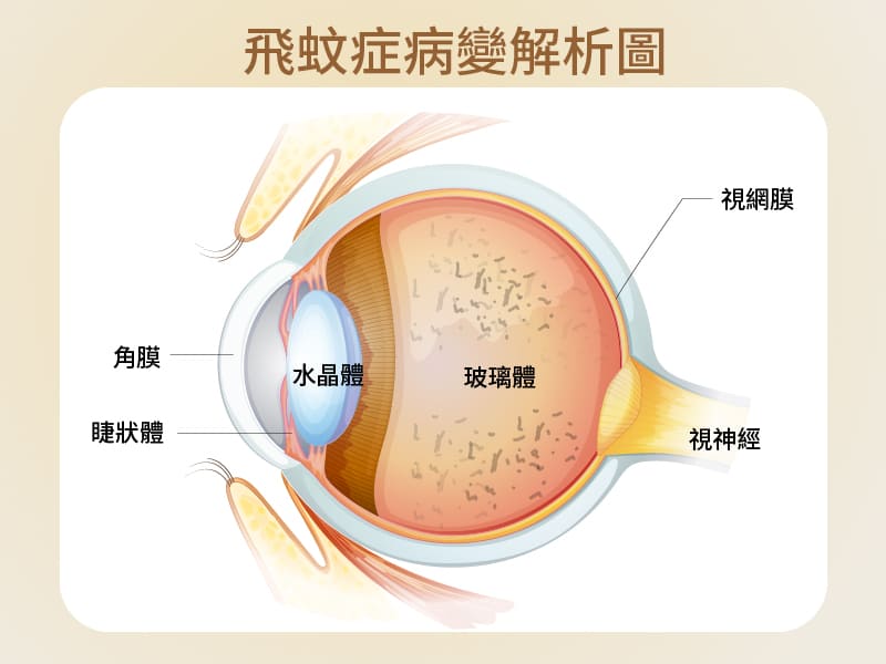
From a medical perspective, floaters are a symptom of vitreous degeneration. The vitreous is a transparent gel-like substance located behind the lens and in front of the retina. Under normal conditions, it fills the entire vitreous cavity, helping to maintain the eye's shape.However, with aging and conditions like myopia, the vitreous liquefies and shrinks, forming cloudy fibers. These fibers float within the vitreous cavity. When light rays refract off these fibers, patients perceive dark spots in their vision—the precursor to floaters.
From a medical perspective, floaters are a symptom of vitreous degeneration. The vitreous is a transparent gel-like substance located behind the lens and in front of the retina. Under normal conditions, it fills the entire vitreous cavity, helping to maintain the eye's shape.However, with aging and conditions like myopia, the vitreous liquefies and shrinks, forming cloudy fibers. These fibers float within the vitreous cavity. When light rays refract off these fibers, patients perceive dark spots in their vision—the precursor to floaters.
Who is most likely to develop floaters?
Floaters typically become more common with age, making older adults more prone to experiencing dark spots in their vision. Beyond age, other factors that may increase the risk of developing floaters include: myopia, a history of eye surgery, eye trauma, or eye inflammation. Clinical medical research indicates that high-risk groups for floaters include:
Middle-aged and elderly people
Individuals with high myopia
Patients with hypertension/diabetes
Undergone eye surgery
Head injury due to impact, such as: car accidents, diving athletes
Other eye problems, such as: eye inflammation
However, most cases of floaters are benign. They can occur due to age-related vitreous degeneration or excessive eye strain. As long as the number of dark spots in your vision doesn't increase and their position remains stable, there's generally no need for excessive concern.
Conversely, if you suddenly experience a large number of dark spots in your vision that are already affecting your field of vision or accompanied by flashes of light, it could indicate vitreous detachment. This condition may cause traction on the retina, leading to retinal tears or even detachment. Such situations require immediate examination to seek treatment options or undergo floater treatment.
Causes of Floaters
The causes of floaters can be broadly categorized into three types: physiological, degenerative, and pathological.
physiological
Approximately 80% of floaters are physiological in nature. Physiological floaters typically occur in individuals under 40 or those who experience prolonged eye strain. Most people notice dark spots in their vision, which are impurities within the vitreous humor. These generally do not affect vision and do not require immediate treatment; they often disappear on their own over time.
Degenerative
As we age, the vitreous humor in the eye gradually degenerates, much like other organs in the body. During this process, the vitreous contracts and forms tiny fibers that float within it. When light enters the eye and refracts off these impurities, it creates the dark spots we see.
pathological
Pathological floaters are caused by eye diseases or systemic vascular conditions. Eye diseases refer to retinal tears or holes that occur due to external traction or degenerative processes, leading to vitreous hemorrhage or even retinal detachment. This can result in vision impairment and, in the most severe cases, permanent blindness.Additionally, systemic vascular diseases such as retinal hemorrhage, diabetes, hypertension, or macular degeneration may also present pathological floaters if not addressed promptly and treated effectively. Patients should seek early intervention or treatment options to manage floaters.
Complications of Floaters — Retinal Detachment/Hole
Although floaters are not a serious condition, retinal detachment caused by floaters is an ophthalmic emergency. One in four patients with floaters may experience vision loss due to retinal detachment or tears. Remember, if you experience any of the following signs of retinal detachment caused by floaters, including:
A large number of dark spots appear in the eyes within a short period of time.
Abnormal Flash
Shadow
Obstructed view
This indicates a potential retinal tear, requiring immediate medical attention and treatment. Otherwise, patients with floaters may suffer permanent vision damage or even blindness.
How to treat floaters?
If your condition is benign floaters, immediate treatment is not necessary. When you notice dark spots in your vision, try moving your eyes to shift the fluid within them, allowing the fibers to drift out of your line of sight. Of course, some individuals find the constant presence of floating dark spots intolerable, as they frequently distract attention and significantly impact mood. In such cases, floater treatment may be considered.
With current technology, the primary methods for treating and improving floaters are laser therapy and vitrectomy surgery.
I. Laser Treatment for Floaters
This treatment method for floaters is suitable for larger, concentrated vitreous fibers. It uses laser energy to break these fibers into smaller fragments, thereby improving and eliminating floater symptoms.Laser treatment is performed under local anesthesia, and patients experience no pain during the procedure. The entire treatment takes only 15 to 20 minutes. However, laser therapy is not suitable for everyone seeking to improve floaters. If the fibers are too scattered or located too close to the macula or lens, the procedure should not be performed to avoid complications such as cataracts or macular damage.
II. Vitrectomy Surgery for Floaters
Vitrectomy surgery involves removing the vitreous gel inside the eye through a small incision and replacing it with a solution to maintain the eye's shape. The procedure typically takes only 10 to 15 minutes. However, vitrectomy does not necessarily eliminate all floaters. If the surgery itself causes bleeding or retinal tears, new floaters may form. Therefore, most physicians do not recommend this surgery for treating floaters.
Methods for Improving and Managing Floaters
Once floaters develop, they are nearly impossible to reverse. Even after laser or surgical treatment, recurrence is possible. What we can do is learn to live with them peacefully. Alternatively, the following methods may help improve floaters and slightly alleviate symptoms.
I. Daily Care for Benign Floaters
Avoid excessive eye strain
Avoid overexerting your eyes during daily activities, especially if you are a heavy user of electronic devices. Take a 5- to 10-minute break every hour to rest your eyes. Use this time to blink frequently and look into the distance to relax your eyes.
Healthy Lifestyle Habits
In daily life, it is recommended to reduce the time spent on your phone, especially avoiding late-night phone use. Ensuring adequate sleep and engaging in regular exercise can also help improve floaters.
Regular follow-up appointments
Never assume that having benign floaters means you can relax. Floater sufferers must undergo regular check-ups to avoid missing other potential conditions and missing the optimal treatment window.
Eye-Protecting Dietary Habits
In daily life, one should increase intake of various vitamins and antioxidant-rich foods through diet. Examples include berries, green or yellow vegetables, carrots, soybeans, milk, and deep-sea fish oil—all of which are dietary approaches that may help improve floaters.
II. Postoperative Care and Maintenance for Malignant Floaters
Topical medication treatment
After surgery, be sure to follow your doctor's instructions for medication. Remember to thoroughly wash your hands with soap before each application to prevent cross-infection.
Improve daily routines
Avoid rapid eye movements and excessive eye strain for one week after surgery. During this period, minimize reading books, using mobile phones, computers, or watching television. Additionally, refrain from strenuous exercise, mountain climbing, scuba diving, or air travel for one month post-surgery, as these activities may stimulate the eyes and slow down the healing process.
Use an eye mask
During the eye recovery period, it is recommended to wear an eye shield at all times to prevent collisions and avoid unintentional rubbing of the eyes while sleeping at night. Ensure the eye shield is clean when in use, and it is advised to wash and disinfect it daily to prevent wound infection caused by bacteria.
Daily diet
In addition to avoiding irritating foods, it is also important to avoid legumes, as these foods can cause nitric oxide in the blood to migrate to the eyes. This leads to gas buildup in the inert gases within the eye, increasing intraocular pressure.
Home Safety
Keep your home clean and tidy to minimize dust and avoid secondhand smoke. During recovery, avoid rearranging furniture or placing items in hallways to prevent collisions or falls in unfamiliar surroundings that could injure your eyes.
Scheduled follow-up appointments
In addition to following your doctor's instructions for regular post-operative follow-up appointments, if you experience any eye discomfort—such as persistent eye pain, sudden blurred vision, nausea, or an increase in the number or size of floaters—be sure to consult your ophthalmologist immediately to prevent the condition from worsening.
How to prevent floaters?
Although we cannot completely prevent the occurrence of floaters, we can slow down the aging of the eyeball and reduce the likelihood of developing floaters through daily eye care.
I. Develop Good Eye Care Habits
Avoid excessive use of electronic devices (take a 5-10 minute break after every hour of screen time).
Work under conditions of sufficient lighting and stable, flicker-free light sources.
Avoid staying up late to reduce strain on your eyes.
II. Consuming Eye-Protective Nutrients
Carotenoids (dark green, deep yellow, and red fruits and vegetables)
Anthocyanins (Plum fruits)
Vitamins (Vitamins A, B complex, C, and E)
Omega-3 fatty acids (fish oil)
III. Properly manage systemic vascular diseases (e.g., diabetes, hypertension)
Maintain a balanced diet and reduce intake of foods high in fat, salt, and sugar.
Regular exercise (jogging, cycling)
Get regular full-body checkups
Although most cases of floaters do not require treatment, they should not be completely ignored. Regular check-ups are essential, and if symptoms worsen, seek medical attention immediately to avoid missing the optimal window for treatment.
#floating dark spots in eyes #improving dark spots in eyes #dark spots on iris #dark spots in eyes
Many seniors experience blurred vision and may attribute it to age-related decline, but have they considered that it could stem from an eye disease?
Eye Disease Symptoms
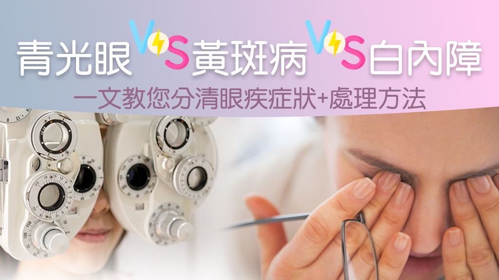
First, determine whether vision loss is sudden or gradual, and whether it affects central vision, the entire visual field, or peripheral vision. Also note if it's accompanied by eye pain. How can we distinguish between macular degeneration, cataracts, and glaucoma? Take cataracts as an example: vision in older adults typically declines gradually and affects the entire visual field. Macular degeneration, conversely, causes sudden, acute vision loss affecting central vision.Glaucoma also gradually affects vision, typically starting with peripheral vision loss.
These three conditions are easily confused, and seniors can check for them through various methods. One method involves using "checkered paper." This grid-patterned paper allows seniors to observe whether straight lines appear curved. Such distortions can indicate the presence of macular degeneration. For other issues, an ophthalmologist's examination is necessary to determine the specific eye condition.
How to treat it?
Diabetic retinopathy typically presents with two complications, primarily caused by oxygen deprivation leading to vascular proliferation. When blood vessels proliferate, these fragile vessels may leak, resulting in vitreous hemorrhage or macular edema. To prevent this, intravitreal injections are commonly administered to inhibit vascular proliferation and thereby reduce oxygen deprivation.
Cataracts are primarily treated through minimally invasive cataract surgery, which involves removing the clouded lens and implanting an artificial lens. Finally, glaucoma can be managed through early intervention to prevent ongoing damage to the optic nerve. There is no cure for glaucoma; treatment focuses on monitoring eye pressure through regular testing and managing it with eye drops, surgery, or laser procedures to slow nerve damage.
Seniors can prevent eye diseases by making lifestyle changes, such as: reducing sugar and salt intake; consuming foods rich in vitamin A, carotenoids, or omega fatty acids for their antioxidant effects; engaging in at least 30 minutes of aerobic exercise daily; and avoiding smoking. Regular eye examinations are essential for early prevention and treatment. Minimizing exposure of the eyes to ultraviolet light is crucial for preventing various eye conditions.
Many doctors use the eyes as a window to observe bodily ailments, especially in patients with chronic conditions. So, which diseases can affect the eyes?
Diseases Affecting the Eyes
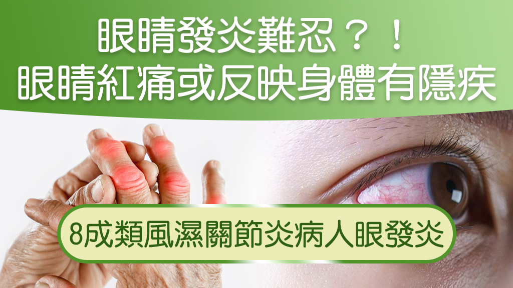
Eye pain, redness, and inflammation are common experiences among older adults. However, if these symptoms persist, we may become concerned about underlying health issues. One such concern is an autoimmune disorder, which can trigger inflammation throughout the body. The eyes are among the organs affected. If left unaddressed or untreated, this can lead to progressive vision loss and even the risk of blindness.
Symptoms and Treatment of Immune System Disorders
Immune system disorders can affect people of all ages, with the most common being rheumatoid arthritis, Sjögren's syndrome, ankylosing spondylitis, or lupus. Taking rheumatoid arthritis as an example, patients may experience lower back pain, hip joint pain, or inflammation symptoms that cause stiffness and soreness in the back upon waking. Due to inflammation, calcification may occur.Joints may fuse together, making it difficult to bend over or reach down to touch one's toes upon waking. Over time, this can significantly impair joint mobility. First-line treatment involves systemic therapy, typically prescribed by rheumatologists in the form of oral anti-inflammatory medications.
First-line treatment typically involves steroids. However, when high-dose steroids or prolonged therapy is required, second-line medications are generally used to mitigate steroid-related side effects. These second-line agents include biologics and immunosuppressants for long-term disease management.
Research indicates that eight out of ten patients will experience at least one episode of eye inflammation in their lifetime. Iritis is one such inflammatory condition that releases cells causing vision loss. These cells bind to proteins within the eyeball and accumulate in the anterior chamber angle, obstructing the drainage of aqueous humor. This leads to increased intraocular pressure, damaging the optic nerve and potentially resulting in glaucoma.
For treatment, blood tests are typically conducted to measure inflammatory markers, and X-rays are taken—such as chest X-rays or lumbar spine X-rays—to check for lung or spinal issues. If problems are found, medication is usually prescribed to control inflammation (specifically, anti-inflammatory steroid eye drops). Additionally, monitoring for side effects and complications is essential, with glaucoma medication used to manage intraocular pressure. Therefore, both ophthalmologists and rheumatologists collaborate to provide comprehensive care and treatment for the patient.
Medicine
Eye drops come in different types. Some are merely lubricating solutions that offer no therapeutic benefit and may even delay proper treatment. Others constrict blood vessels but do not control inflammation, potentially creating a false impression of ongoing improvement. Finally, if the treatment dosage is insufficient or the duration of use is inadequate, it can delay proper care and lead to irreversible damage to the eyes.
COVID-19
Conjunctivitis often accompanies respiratory infections such as influenza or viral colds, and it is also one of the symptoms of COVID-19. The most common symptom of conjunctivitis is redness and inflammation of the conjunctiva covering the white part of the eye. Research shows that among 30 COVID-19 patients examined during hospitalization, one exhibited conjunctivitis symptoms, indicating a close association.
To prevent COVID-19, first and foremost, we must wash our hands frequently with soap and water for at least 20 seconds. Additionally, we need to maintain a proper social distance of 1.5 meters and ensure adequate ventilation to protect ourselves from the virus.