Symptoms and Causes of Presbyopia
Presbyopia is a normal physiological phenomenon. Its primary characteristic is a decline in near vision, particularly when reading or performing close-up tasks. Presbyopia is a vision problem that develops with age; symptoms typically begin to appear around the age of 40. It is caused by the gradual deterioration of the ciliary muscles and the lens, resulting in a slower ability to focus.
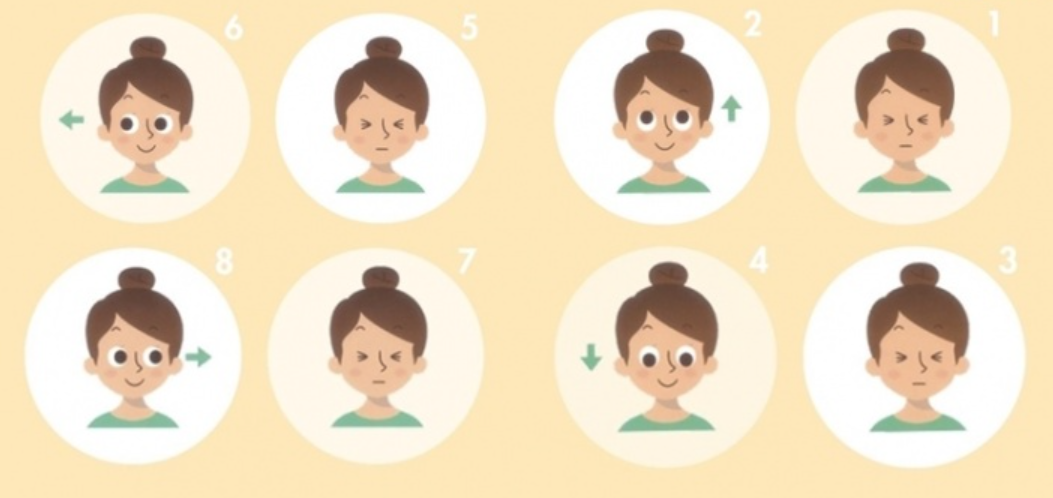
8 Common Symptoms of Presbyopia
Although presbyopia isn’t considered a serious eye condition, the degree of presbyopia tends to worsen with age, and its symptoms become increasingly noticeable, which can significantly impact daily life and work efficiency. Here are some symptoms that may indicate you have presbyopia:
- When reading or looking at a phone, I have to hold it more than 40 centimeters away from my eyes to see the text clearly.
- Eyes tend to feel tired, dry, or sore.
- It is difficult to see nearby objects in dimly lit environments.
- When I look at something in the distance and then shift my gaze to something closer, it feels like the focus is slower
- You need to blink frequently or rub your eyes to try to adjust your focus.
- I have less time to read, so I need more time to rest.
- Blurred or fuzzy vision, especially when looking at things up close.
- Performing detailed work, such as intricate painting or crafting, requires more time and effort.
2 Ways to Self-Test for Presbyopia
In fact, the early symptoms of presbyopia are not very noticeable, and many people mistake them for eye strain or temporary blurred vision caused by overuse. Below are two methods you can use at home to check if you have developed presbyopia. However, these are not professional diagnostic methods. If you notice symptoms of presbyopia, we recommend that you visit a professional eye care center for a comprehensive examination and to develop a treatment plan tailored to your needs.
Banknote Testing
- Take out a bill and place it where you can see it most clearly.
- Then focus on the fine print on the banknote.
- Look away from the banknote and focus on an object about 2 to 3 meters away for approximately 5 seconds.
- Then look back at that line of small print and see how many seconds it takes to make out the text.
Focusing within 1 second is considered normal; Taking longer than 1 second indicates a risk of developing presbyopia; Taking over 3 seconds confirms the presence of presbyopia.
Symptoms and Screening for Presbyopia
- When looking at objects, I find myself wanting to take off my glasses—yet without them, I see more clearly.
- Feeling that your home or office is dimmer than before
- Often misread numbers or text
- I often feel eye strain.
- Stiffness and soreness in the shoulders and neck
- Frequently frowns, experiences dizziness
These are all common signs of presbyopia. If you experience three or more of these symptoms, you may have presbyopia. We recommend consulting an ophthalmologist as soon as possible to get professional advice and assistance.
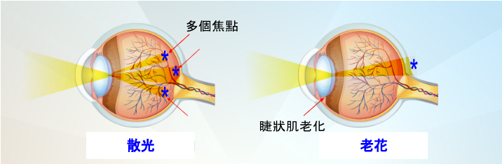
Causes of Presbyopia
The primary cause of presbyopia is the gradual loss of flexibility and elasticity in the ciliary muscles and lens of the eye, which prevents the eye from accurately adjusting its focus. This leads to a decline in near vision, resulting in difficulty seeing objects up close. Presbyopia is typically age-related, but it can also be influenced by other factors.
In addition to natural aging, the following factors may also contribute to the development of presbyopia:
- Genetic factors: Genetics may influence a person’s susceptibility to presbyopia and the age at which it develops.
- Chronic eye strain: Prolonged exposure to electronic screens or other close-up work may cause presbyopia symptoms to appear at a young age.
- Environmental factors: Harsh environmental conditions, such as intense ultraviolet radiation or pollutants, may also accelerate the progression of presbyopia.
How to Prevent and Treat Presbyopia
Almost everyone will eventually experience presbyopia. Although there is currently no proven method to prevent it, there are ways to slow its progression, such as maintaining good eye care habits, giving your eyes adequate rest, and consuming plenty of eye-nourishing nutrients.
If you start noticing signs of presbyopia, there’s no need to worry too much. You can choose from various options to address presbyopia, such as reading glasses, reading contact lenses, LBV laser vision correction, or Presbymax laser surgery.
In summary, presbyopia is an age-related vision condition. To slow the progression of its symptoms, it is important to maintain good eye health on a daily basis and undergo regular eye exams. Early intervention and treatment are essential for effectively preserving good vision and maintaining quality of life.