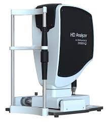
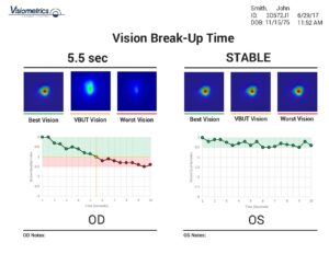
The HD Analyzer is a sophisticated instrument capable of quantifying light scattering within the eye, thereby providing an objective measure of visual quality. This is particularly useful for evaluating cataracts, as it reveals the severity of the cataract and its impact on vision. It also enables an objective assessment of dry eye syndrome by measuring the stability of the tear film, which plays a key role in tear production.By comparing the stability of the tear film over time with ocular scattering, the device assesses the impact of dry eye on visual quality. This helps rule out abnormal tear films that cause vision loss and can also be used to monitor a patient’s response to dry eye treatment.
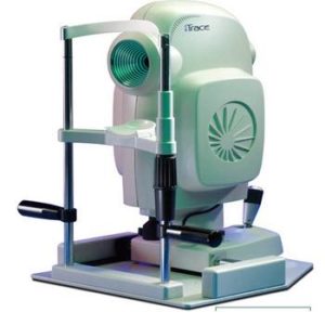
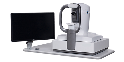
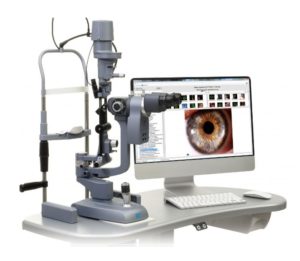
iTrace
It offers a 5-in-1 tool for eye care that analyzes comprehensive visual function. Using a single, easy-to-use device, it measures a patient’s wavefront, corneal topography, pupil size, refraction, and corneal curvature, thereby providing a comprehensive assessment of visual quality.
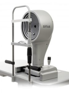
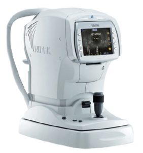
Sirius +
Corneal topography combines the Platt-Dado disc topography with Scheimpflug tomography of the anterior segment to provide information on the thickness, height, curvature, and refractive power of both corneal surfaces within a 12-mm diameter. All biometric measurements of the anterior chamber are calculated using up to 100 high-resolution corneal sections.
AngioVue OCT
Using a non-invasive, non-fluorescent imaging technique, this technology rapidly assesses a patient’s microvascular system and can be performed by trained technicians in a matter of seconds. With AngioVue, you can image patients as needed to closely monitor disease progression and treatment response.
Optovue Epithelial Map
The proprietary Epithelial Thickness Map (ETM) is the first non-contact quantitative measurement of the corneal epithelium and stroma, helping you assess early changes in the ocular surface to aid in the diagnosis of corneal diseases. A quick two-second scan identifies areas of thickening or thinning associated with dry eye disease, keratoconus, or prior refractive surgery.Change analysis allows you to track changes in ETM mapping over time, enabling you to evaluate the efficacy of cross-linking or dry eye treatments. For refractive surgery patients, ETM provides reliable risk analysis by quantifying epithelial and stromal thickness, which can inform surgical planning or help you assess refractive surgery outcomes.
OSA Dry Eye Test
OSA effectively integrates the following comprehensive examinations to provide users with dry eye syndrome reports:
Meibomian Gland Imaging
Demodex Imaging
Imaging of Blepharitis
Tear Breakup Time (NIBUT)
Assessment of the Sebum Layer
Tear Level
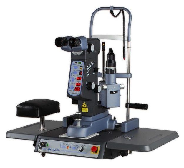
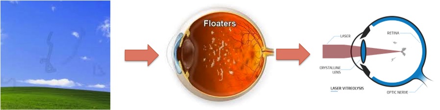
Laser machine
Laser Treatment for Retinal Holes
Laser Removal of Glass Impurities
Laser treatment for posterior capsule opacification
A macular tomography scan displays a cross-sectional image of the macular region.
Examination of various macular conditions, such as macular edema, macular holes, age-related macular degeneration, diabetic retinopathy, and epiretinal membranes.
Optical Coherence Tomography (Glaucoma)
Optic nerve head thickness measurement and CT scan
Measure the thickness of the retinal nerve fiber layer and the cup-to-disc ratio of the optic disc
SLIT LAMP
Examine the conjunctiva, anterior chamber, iris, eyelids, lens, retina, macula, optic nerve, and other structures of the eye.
High-resolution digital images, still photographs, and video recordings help patients and their families understand the condition.
A slit lamp typically consists of two parts: a light source and a binocular microscope—that is, an illumination system and an observation system.
A light source refers to a device whose beam width can be adjusted horizontally and vertically to meet illumination requirements; simultaneously, a binocular microscope focuses on the same point or plane illuminated by the light source, which is also the confocal process between the two components.
Examine the eyelids, conjunctiva, iris, anterior chamber, lens, and retina using a slit-lamp microscope.
Intraocular Pressure and Corneal Curvature Measuring Device
Capable of measuring basic refraction, accommodation, and fundus images
3D Auto Focus and Automatic Measurement
VISUAL FIELD Visual Field Tester
Measure the sensitivity of different areas of the retina to light spots under the instrument’s fixed background illumination.
Visual field testing is primarily used to screen for glaucoma and other eye conditions that cause visual field defects. It helps doctors diagnose and monitor the condition and determine the appropriate treatment plan.
Lenstar Intraocular Lens Measuring System
It can precisely measure the patient’s corneal curvature, axial length, and anterior chamber depth to accurately calculate the power of the intraocular lens used in cataract surgery.
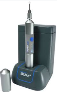
Treatment of Anterior Blepharitis: AB Max™
AB Max™ offers forward and reverse functions, and its patented PULSE mode is specifically designed to remove even the most stubborn crusts and debris while gently massaging the anterior eyelid margin to improve treatment outcomes for patients.
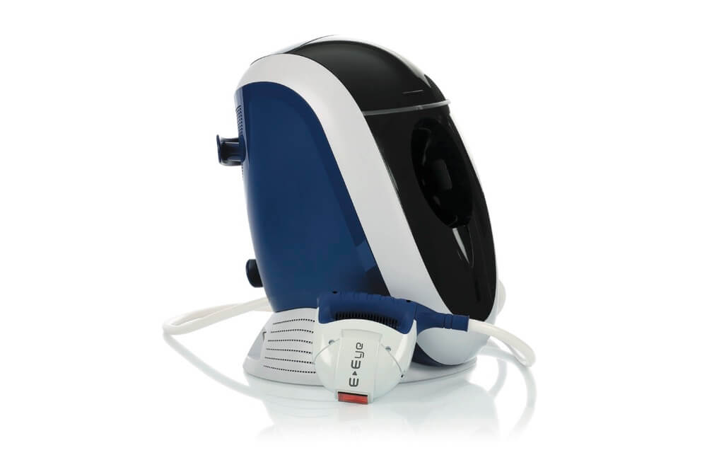
Intense Pulsed Light (IPL)
E-Eye is a device that generates multi-wavelength pulsed light (Intense Pulsed Light, or IPL) to directly treat the meibomian glands in patients with dry eye. Meibomian gland dysfunction (MGD) is a primary cause of dry eye discomfort. Treatment with E-Eye over several sessions stimulates the function of these oil-producing glands, restoring the ocular surface lipid layer. E-Eye delivers uniformly spaced light pulses in the form of a calibrated pulse sequence. The energy, spectrum, and duration are calculated to stimulate the meibomian glands.
It helps patients with dry eye syndrome by reducing abnormal blood vessels (telangiectasia) around the eyelids that secrete inflammatory chemicals, improving meibomian gland function, and reducing the bacterial load around the eyes. The end result: more tears, healthier tear composition, and fewer dry eye symptoms!
The LLLT mask utilizes medical-grade LEDs for photomodulation, a form of photobiomodulation technology that stimulates the production of ATP (adenosine triphosphate) by emitting light at specific wavelengths. It also significantly heats the eyelids to the temperature recommended for periorbital thermal therapy. The combination of increased cellular activity triggered by endogenous heat and external thermal heating helps stimulate glandular normalization. Indications for this treatment include meibomian gland dysfunction (MGD), dry eye, blepharitis, and styes.