有散光又有老花,LBV 激光矯視可以一次過矯正?
戴咗幾十年眼鏡,而家唔單只睇遠要靠鏡,睇近都開始困難,度數單上寫住「有散光、有老花」,眼鏡愈換愈複雜,但視力就係唔夠清晰。想做 LBV 老花激光矯視,但有散光係咪會影響手術?係咪散光要另外處理,先至可以做老花?
其實,LBV 在設計切削方案時,可以同時將散光度數計算在內,令散光、老花喺同一次手術中一併矯正。但散光嘅程度、類型,以及角膜形態,都係決定可否一次過處理嘅關鍵因素。
散光係點嚟?點解會同老花一齊出現?
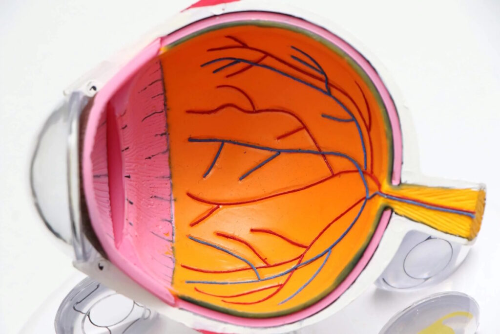
散光係因為角膜表面唔夠圓滑,形狀略似橄欖球而非正圓,令光線進入眼睛後無法集中喺同一焦點,視物出現模糊或重影。散光可以單獨存在,亦可以同近視、老花同時出現喺同一個人身上。
隨住年齡增長,晶狀體彈性退化引發老花,與此同時,角膜形態亦可能因長期磨損而輕微變化,令本來輕微嘅散光變得更為明顯。因此,50 歲後同時有散光同老花係非常普遍嘅情況,兩者加起來令配鏡難度大增,漸進鏡嘅適應亦更困難。
另一個唔少人忽略嘅事實係:散光對視力嘅影響,在明亮環境同昏暗環境下感受差異明顯。白天光線充足時,瞳孔較細,散光影響相對輕微;夜晚或燈光昏暗嘅環境,瞳孔放大,散光令影像更容易出現重影或光暈,睇街燈、車燈時尤為明顯。如果你發現夜間視力特別差、睇光源周圍有拖尾或星芒,散光可能係原因之一。
LBV 可以同時矯正散光同老花?
LBV(Laser Blended Vision)嘅手術平台在設計雙眼切削方案時,可以將散光度數整合入矯正計劃之中。主視眼在矯正遠距視力嘅同時,同步處理散光;非主視眼在設定近距焦點嘅同時,亦一併矯正散光。手術完成後,理想情況係散光同老花兩個問題都得到處理,日常生活毋須再依賴眼鏡。
散光嘅矯正效果取決於幾個條件:散光度數嘅高低、散光軸向係咪規則,以及角膜形態嘅穩定性。一般而言,規則性散光(Regular Astigmatism)對激光矯正嘅回應較為理想,不規則散光(Irregular Astigmatism)則需要更仔細嘅術前評估,部分情況下可能需要先處理角膜問題。
💡 散光程度對 LBV 嘅影響
輕至中度散光(通常 3.00D 或以下)在大部分情況下可於 LBV 手術中一併矯正。散光度數較高或角膜形態不規則者,需要額外評估角膜地形圖,確認切削設計係咪可以安全涵蓋所有度數。
有散光同老花,LBV 術前需要評估什麼?
術前評估係確保 LBV 能夠安全有效處理散光同老花嘅必要步驟。評估內容通常包括:
| 評估項目 | 目的 |
|---|---|
| 角膜地形圖 | 確認散光係規則性或不規則性,評估角膜形態是否穩定 |
| 角膜厚度測量 | 確保角膜有足夠厚度支撐同時矯正散光、老花所需嘅切削量 |
| 主視眼測試 | 確定哪隻眼主責遠距,哪隻眼負責近距,制定 LBV 雙眼方案 |
| 散光軸向確認 | 精確記錄散光方向,確保切削設計對準正確軸向 |
如果散光程度偏高或角膜地形圖顯示不規則形態,醫生可能建議先以其他方式穩定角膜,或評估是否適合 LBV 以外嘅矯正方案,例如植入式隱形眼鏡(ICL)或換晶體手術。
LBV 矯正散光同老花後,生活有咩改變?
對於長期同時戴近視鏡、散光鏡同老花鏡嘅人士,LBV 術後最直接嘅改變,係唔再需要頻繁換眼鏡。駕駛時睇遠清晰、對比度提升;閱讀時毋須拉遠手機或書本;散光引發嘅夜間重影或星芒感,在術後亦通常有所改善。
對於 45 至 60 歲仍在工作嘅人士,例如需要長時間對住電腦或文件,或者經常要切換遠近視距嘅工作環境,LBV 同時矯正三個問題(近視、散光、老花)後,可以減少視覺疲勞及換鏡麻煩,工作效率亦可能因此有所提升。
常見問題 FAQ
Q1:有散光同老花,LBV 真係可以一次矯正兩個問題?
大部分情況下可以。LBV 嘅激光切削設計可整合散光同老花嘅矯正度數,令兩者在同一次手術中一併處理。但能否一次矯正、矯正效果係點,取決於散光度數、散光類型及角膜厚度,需術前評估確定。
Q2:散光度數幾高先係 LBV 嘅限制?
一般而言,規則性散光約 3.00D 或以下,在多數情況下可與老花同時矯正。散光度數較高者,需要評估角膜剩餘厚度是否足夠,以及散光軸向是否適合激光切削設計。具體上限因個人角膜條件而異,術前評估可給出明確答案。
Q3:散光係規則性定不規則性,點知?有咩分別?
規則性散光係角膜兩個主軸呈 90 度垂直,光學矯正較為直接;不規則散光則係角膜形態複雜,無法用簡單嘅軸向描述,常見於曾受過傷或有圓錐角膜嘅患者。規則性散光對 LBV 激光矯正嘅回應通常較理想,不規則散光則需更詳細評估。
Q4:做完 LBV 後,散光同老花係咪都唔會再有?
LBV 可有效矯正手術時嘅散光及老花度數。散光矯正後通常較為穩定,因為角膜形態改變係永久性嘅。老花方面,由於屬於持續退化嘅生理現象,日後度數仍可能輕微增加,部分人若干年後睇近時可能需要輕度老花鏡。
Q5:有散光嘅人做 LBV,術後適應期係咪更長?
散光矯正後,視覺質素通常在短期內已有明顯改善。LBV 本身需要大腦適應雙眼景深融合,適應期一般係數天至數週。有散光嘅人並不代表適應期一定更長,但每個人嘅適應進度有所不同,術前了解適應過程有助建立合理期望。
Q6:我有輕微散光,驗光師話唔需要特別矯正,LBV 做唔做得?
輕微散光(如 0.50D 或以下)在日常生活中影響通常不大,LBV 手術計劃可以選擇一併矯正或不特別針對處理,視乎個人視力需求同角膜條件。術前與醫生討論嘅時候,可以提出呢個問題,了解矯正散光對你整體視力嘅影響。
Q7:散光加老花,係咪換晶體比 LBV 更好?
兩者各有適應條件。LBV 係在角膜進行激光切削,適合角膜條件良好、無白內障嘅人士;多焦點散光晶體則係眼內手術,適合同時有散光、老花及初期白內障嘅人士。選擇哪條路線,需要根據個人眼部條件、年齡及視力需求由醫生評估建議。散光程度較高嘅人士,換晶體方案有時反而更能一步處理所有度數問題,值得一同納入評估考量。
有散光又有老花,LBV 激光矯視可以一次過矯正? Read More »