青光眼類型

Recognizing different types of glaucoma: What are the differences between open-angle, closed-angle, congenital, and normal-pressure glaucoma?

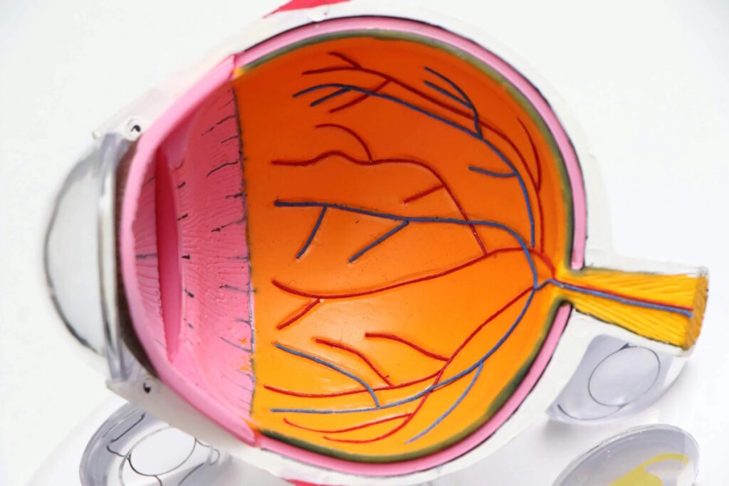
Glaucoma is not a single disease but a group of eye conditions that damage the optic nerve. Although elevated eye pressure is the main risk factor, some patients may still develop glaucoma even if their eye pressure is normal. Common types include open-angle glaucoma, closed-angle glaucoma, congenital glaucoma, and normal-tension glaucoma, each with different causes and symptoms.

Angle-shaped glaucoma

Primary angle-closure glaucoma is the most common type of glaucoma, accounting for about 6 to 8 percent of cases. The anterior chamber appears open, but as the function of aqueous humor drainage in the eye gradually declines, the drainage is obstructed, causing chronic elevated eye pressure and long-term pressure on the optic nerve. Age, family history, high myopia, diabetes, and hypertension all increase the risk of developing the disease.

Early open-angle glaucoma has almost no obvious symptoms. The central vision usually remains normal. Patients are not easily aware of it. As the condition worsens, the peripheral vision gradually narrows, and they may feel a lack of peripheral vision, have difficulty walking, and see things as if a part of them is blocked; however, many people do not seek medical attention until the visual impairment is significantly impaired.

Angle-closure glaucoma

Primary angle-closure glaucoma is more common in Asians, mainly due to the anatomy of the eyeball: the anterior chamber is abnormally narrow or closed, and the lens is thicker, which prevents aqueous humor from flowing properly into the trabecular meshwork and causing a significant increase in eye pressure within a short period of time, leading to angle-closure glaucoma.

Symptoms are very noticeable during an acute angle-closure glaucoma attack, including severe eye pain, red eyes, sudden blurred vision, rainbow-colored halos when looking at lights, severe headaches, nausea, and even vomiting. It is an emergency for ophthalmology, requiring immediate medical attention to prevent rapid damage to the optic nerve. In chronic angle-closure glaucoma, the angle gradually closes, with more subtle symptoms, such as occasional eye swelling and slight blurred vision. Over time, this can also narrow the field of view.

Congenital glaucoma

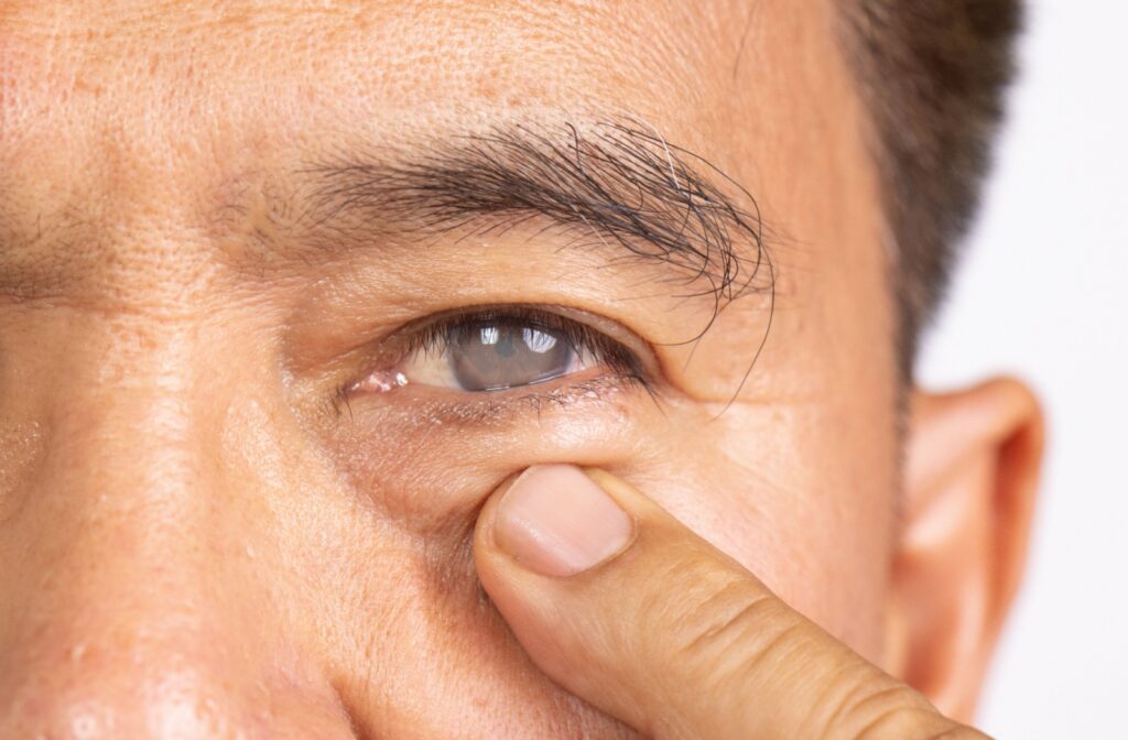
Congenital glaucoma mostly occurs in infants or young children. Typical symptoms include the eyeball being larger than that of a normal infant, the cornea being cloudy or grey-white, sensitivity to light, frequent tearing of the eyes, and frequent rubbing of the eyes with hands. This may lead to delayed visual development.

The cause of congenital glaucoma is structural abnormalities in the drainage system during development, leading to elevated intraocular pressure from an early age. This is related to genetic or developmental issues in the eye during fetal development, and is usually diagnosed within the first three years of life. Since children may not be able to express discomfort, parents should arrange for an eye examination as soon as possible if they notice abnormal eye appearance or persistent light sensitivity in their child.

Normal ocular pressure glaucoma

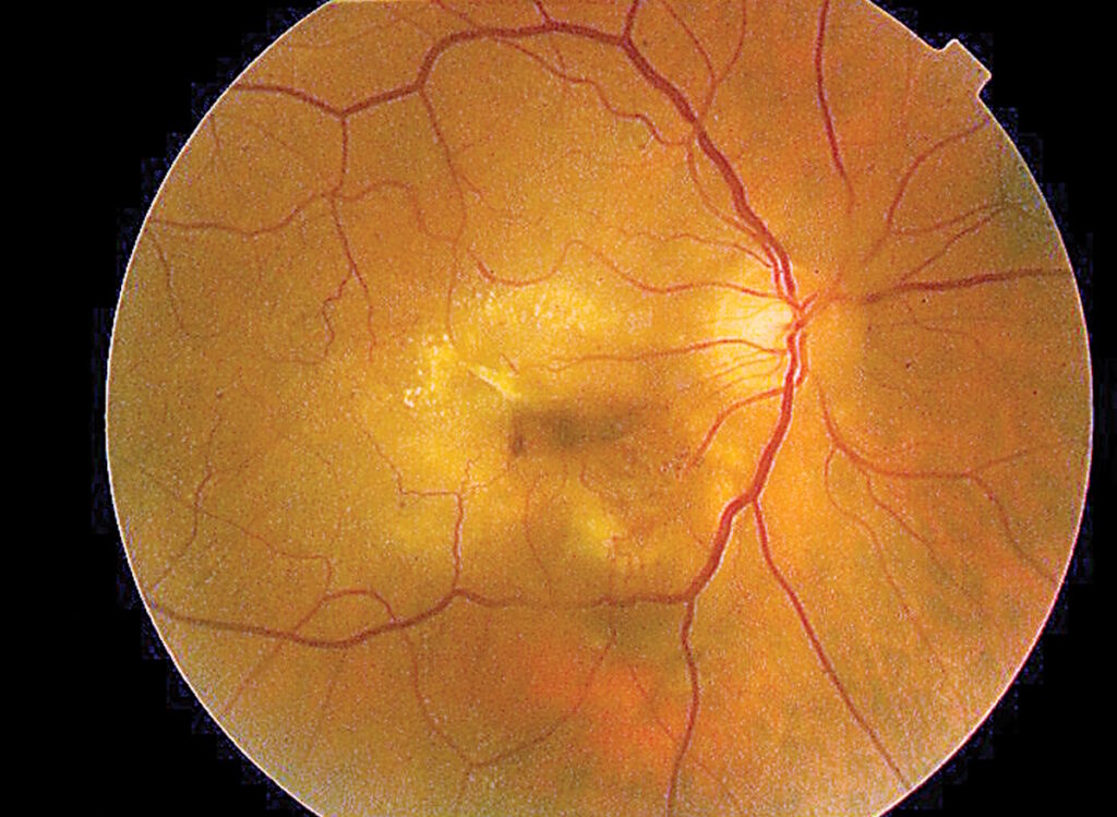
The characteristic of normal ocular pressure glaucoma is that the eye pressure is within the normal range, but the optic nerve still shrinks. The optic nerve gradually loses its function. The medical community believes that this is a similar condition to angle-closure glaucoma; however, some patients have particularly sensitive optic nerves to eye pressure, or the blood flow to the optic nerve is poor, and even if the eye pressure is not particularly high, it can still be damaged.

The symptoms are similar to those of chronic open-angle glaucoma; in the early stages, there are almost no self-conscious symptoms; the patient is usually only discovered during a detailed visual nerve examination, OCT scan, and visual field analysis. If treatment is not initiated in time, the field of vision will gradually narrow, and in severe cases it can even lead to blindness.

Can Glaucoma and Cataracts Be Caused by Each Other?

Cataracts and Glaucoma are common eye diseases, but the pathological mechanisms are not the same;CataractThe main effect of crystalline lens opacity on vision is blurred vision; cataracts are damage to the optic nerve. They do not directly cause each other; however, factors such as age, diabetes, and long-term use of steroids can both increase the risk of cataracts and are also associated with some types of cataracts, so they often occur simultaneously in older adults.

Additionally, severe cataracts thicken the lens, potentially making the anterior chamber increasingly narrow, increasing the risk of developing acute angle-closure glaucoma; while some glaucoma surgeries can affect the lens, increasing the risk of developing cataracts in the future. Therefore, if you already have one of these conditions, you should schedule regular checkups with your doctor for any other problems as well.

Dr. Tong Wenyue, an ophthalmologist, said, "In cases of eye diseases caused by glaucoma, patients usually have high eye pressure, so they check whether the optic nerve shows signs of glaucoma. If there are signs of glaucoma, the pupil will expand, and the doctor will perform more detailed examinations, such as using a visual field chart or optical coherence tomography to check whether the visual field has narrowed and whether the optic nerve has become thinner due to damage."

How to prevent glaucoma?

Although Glaucoma cannot be completely prevented, it can be reduced through regular check-ups and maintaining healthy lifestyle habits. Dr. Tong Wenjie, an ophthalmologist, recommends:

  • People aged 40 or older should undergo a comprehensive eye examination once a year, especially those with a family history of glaucoma, high myopia, or diabetes.
  • Controlling blood pressure, blood sugar and cholesterol levels, managing cardiovascular and metabolic diseases, and reducing damage to the optic nerve blood flow.
  • Avoid smoking and excessive drinking, maintain adequate sleep and a balanced diet, and consume more antioxidant and Omega‑3 fatty acid foods.
  • When using electronic products, take a 20–30-minute break every 20–30 minutes to reduce excessive eye fatigue and avoid looking at the phone for long periods of time in extremely dark environments.

Cataract causes permanent damage to the optic nerve in the eye. Once the vision is lost, it cannot be restored. Therefore, “early detection and early treatment” is the key to protecting vision. Even if there are no symptoms, it is recommended to undergo a comprehensive eye examination regularly, especially for high-risk individuals, and to visit the eye doctor for regular check-ups as recommended by the eye doctor.
Common problems

Recognizing different types of glaucoma: What are the differences between open-angle, closed-angle, congenital, and normal-pressure glaucoma? Read More »

年青人 黃斑病變

Do all young people develop macular degeneration?

Many people think macular degeneration is an eye disease that only affects older adults. However, young people are not completely at risk. Although macular degeneration is more common in older adults, people with nearsightedness have their eyeball stretched, and the retina and macular tissue may be pulled and thinned, increasing the chance of near-sighted macular degeneration. If a person has high myopia, they are more likely to develop macular degeneration.

Dr. Tong Wenjie, an ophthalmologist, explained that the macular area, located in the center of the retina, is the most densely populated area of photoreceptors, which dominate and control our central vision. In essence, it is like the film in a camera. Once the macular area is damaged, it can lead to severe visual impairment. The loss of central vision can lead to deformation and even scarring, and in some cases, blindness.

年青人 黃斑病變

Near-sightedness and the macular area are related

The relationship between macular degeneration and the pathological changes that can be caused by high myopia is profound. The deeper the nearsightedness and the longer the eyeball, the more likely the macular and peripheral retina may be damaged by prolonged stretching, even leading to holes, atrophy, or abnormal blood vessel growth. This type of disease can cause a decline in central vision and may also result in distorted images or shadows blocking out the view. For those with myopia of 600 degrees or more, or a family history of macular disease, heightened vigilance is even more necessary.

Causes of the trend towards youthification

Age remains the main risk factor, but if young people have high myopia and lack the habit of protecting their eyes, the risk of macular degeneration may occur earlier. The aging of macular degeneration is closely related to modern lifestyle habits.

  • Long-term use of mobile phones, computers, and tablets puts the eyes under a high load for a long time, which can easily exacerbate the development of myopia.
  • Reduced outdoor activities, insufficient sunlight, lack of sleep, and too close a distance between the eyes can all cause myopia to deepen.
  • Imbalanced diet, a lack of antioxidant nutrients, and habits such as smoking can also weaken the health of the retina.

Prevention of macular degeneration

To prevent macular degeneration, the key is to monitor and protect the eyes early. People with high myopia should undergo regular comprehensive eye examinations, especially fundus examinations, to detect macular or retinal abnormalities early. In daily life, you can increase your intake of foods containing lutein, zeaxanthin, vitamins C, E, and zinc, such as deep green vegetables, fish, and orange fruits. When going out, wear sunglasses with UV protection to reduce the harmful effects of ultraviolet rays; also quit smoking, control blood pressure, blood lipids, and blood sugar, and avoid prolonged near-sighted use of the eyes. These methods can effectively reduce the risk of macular degeneration and slow its progression.

Laser vision correction for nearsightedness

SMILE Laser VisionThe main role is to correct nearsightedness by changing the curvature of the cornea and improving the position of light focus, thereby reducing or eliminating the nearsightedness. It is suitable for patients with nearsightedness within 1,000 degrees and within 500 degrees of farsightedness. It is important to note that laser vision correction cannot directly treat macular degeneration; those who were previously considered to have high myopia still have a risk of macular degeneration. To effectively treat macular degeneration, it depends on whether it is early dry or late wet macular degeneration. For more details, please refer toMethods for treating macular degeneration。

As the number of people with myopia increases and eye-care habits change, the trend towards macular disease is worth paying attention to. The earlier people establish the habit of checking, protecting their eyes, and controlling their refractive error, the better they can protect the health of the macula and avoid damage to central vision. For people with severe myopia, regular eye examinations and professional assessments can effectively reduce the risk.

Common problems

Do all young people develop macular degeneration? Read More »

有散光又有老花,LBV 激光矯視可以一次過矯正?

Can an LBV laser correction treat both presbyopia and hyperopia at the same time?

Having worn glasses for decades, I now have to rely on glasses not only for distance vision but also for near vision. The prescription on the lens says "with presbyopia," and my glasses are getting more complicated. But my vision isn't clear enough. I want to undergo laser refractive surgery for presbyopia, but will having presbyopia affect the surgery? Does presbyopia need to be treated separately before I can undergo refractive surgery for presbyopia?

In fact, when designing the cutting plan, LBV can simultaneously calculate the scatter level, allowing the scatter and presbyopia to be corrected simultaneously during the same surgery. However, the degree, type, and shape of the cornea are key factors that determine whether the treatment can be performed at once.

What causes presbyopia? Why does it often occur along with cataracts?

Strabismus is caused by the cornea having an irregular surface that is more like an olive rather than a perfect circle, which prevents light from focusing on a single point when it enters the eye. This can result in blurred vision or double vision. Strabismus can occur alone or in combination with myopia and presbyopia in the same person.

As age progresses, the elasticity of the crystalline lens deteriorates, leading to presbyopia. At the same time, the shape of the cornea may also undergo slight changes due to prolonged wear, making the already slight astigmatism more noticeable. Therefore, having both presbyopia and cataracts after age 50 is very common; together, these conditions make lens fitting more difficult and the adaptation to progressive lenses more challenging.

Another fact that many people overlook is that the effect of chromatic aberration on vision varies significantly between bright and dim environments. When there is plenty of light during the day, the pupil is smaller, and chromatic aberration affects vision relatively slightly; at night or in dimly lit environments, the pupil enlarges, making it easier for chromatic aberration to cause heavy shadows or halos on the image. This is especially noticeable when looking at streetlights or car headlights. If you notice particularly poor night vision and see trailing lines or star-like patterns around the light source, chromatic aberration may be one of the causes.

Can LBV simultaneously correct farsightedness and presbyopia?

The LBV (Laser Blended Vision) surgical platform can integrate the refractive error into the correction plan when designing the double-eye cutting scheme. While correcting distance vision, the main eye simultaneously addresses refractive errors; while setting the near focus, the non-main eye also corrects refractive errors simultaneously. After the surgery is completed, the ideal situation is that both the refractive error and presbyopia are addressed, and daily life no longer requires the use of glasses.

The effectiveness of refractive correction depends on several factors: the degree of refractive error, whether the axial direction of the refractive error is regular, and the stability of the corneal shape. Generally speaking, regular refractive error (Regular Astigmatism) responds better to laser correction, while irregular refractive error (Irregular Astigmatism) requires more detailed pre-operative evaluation and may require addressing the corneal problem first in some cases.

💡 The effect of chromatic aberration on LBV

Mild to moderate refractive errors (usually 3.00D or below) can usually be corrected during an LBV procedure. Those with higher refractive errors or irregular corneal morphology require additional evaluation of the corneal topography to confirm that the surgical design can safely cover all refractive values.

💬 Book an eye assessment

There are both presbyopia and hyperopia. I would like to know if it is suitable to undergo LBV. Please feel free to contact us for more information.

WhatsApp inquiry

There are both presbyopia and hyperopia. What should I evaluate before the LBV procedure?

Preoperative evaluation is a necessary step to ensure that LBV can safely and effectively treat presbyopia and hyperopia. The evaluation usually includes:

Evaluation items Purpose
Corneal topography map Confirm whether the astigmatism is regular or irregular, and assess whether the corneal shape is stable.
Corneal thickness measurement Ensures the cornea has sufficient thickness to support the cutting amount required to correct presbyopia and farsightedness simultaneously.
Main visual acuity test Determine which eye is responsible for distance vision and which eye is responsible for near vision; develop an LBV dual-eye program
Radial axis verification Accurately record the direction of scatter light to ensure that the cutting design is aligned with the correct axis.

If the degree of astigmatism is high or the corneal topography shows irregular shapes, the doctor may recommend first stabilizing the cornea with other methods, or assess whether an alternative treatment method other than LBV is suitable, such as implanted contact lenses (ICL) or cataract surgery.

After LASIK and PRK, what changes does life bring about?

For individuals who wear both reading glasses, distance glasses, and bifocal glasses simultaneously on a long-term basis, the most immediate change after the LBV procedure is that they no longer need to frequently change their glasses. When driving, they can see clearly at a distance and the contrast improves; when reading, they no longer need to strain to see the phone or book clearly; and the nighttime glare or star-like light caused by presbyopia usually improves after the procedure.

For people aged 45 to 60 who are still working, such as those who spend long periods of time working with computers or documents, or who frequently need to switch between near and far vision environments, LBV can reduce visual fatigue and the hassle of changing glasses while correcting three problems (myopia, astigmatism, and presbyopia), and work efficiency may also improve as a result.

Common questions FAQ

Q1: Can LBV correct both my presbyopia and my farsightedness at once?

In most cases, it is possible. The LBV laser cutting design can integrate the correction of presbyopia and hyperopia, allowing both to be treated simultaneously during one surgery. However, whether correction can be achieved at one time and the effectiveness of the correction depend on the degree of presbyopia, the type of hyperopia, and the corneal thickness; this must be determined before the procedure.

Q2: What is the maximum dispersion number for an LBV?

Generally, regular astigmatism is approximately 3.00D or less, and in most cases it can be corrected with presbyopia. For those with higher astigmatism levels, it is necessary to assess whether the remaining corneal thickness is sufficient and whether the axial astigmatism is suitable for the laser cutting design. The specific upper limit varies depending on the individual corneal conditions; a pre-operative evaluation can provide a clear answer.

Q3: Is diffuse light regular or irregular? How do you know? What are the differences?

Regular irregular astigmatism occurs when the two main axes of the cornea are at 90 degrees perpendicular to each other; optical correction is more straightforward. Irregular irregular astigmatism is more complex in its corneal morphology and cannot be described in simple axial terms; it is common in patients who have undergone trauma or have a conical cornea. Regular irregular astigmatism usually provides a more ideal response to LBV laser correction; irregular irregular astigmatism requires more detailed evaluation.

Q4: After doing LBV, will the scattered light and presbyopia no longer be a problem?

LBV can effectively correct presbyopia and hyperopia during surgery. After correcting presbyopia, the degree of vision usually stabilizes because the corneal shape changes are permanent. As for hyperopia, since it is a persistent physiological phenomenon, the degree of vision may still slightly increase in the future. For some people, near vision may require mild reading glasses after a few years.

Q5: People with scattered light vision should undergo LBV. Is the recovery period after the procedure longer?

After refractive lens correction, visual quality usually shows significant improvement within a short period of time. The initial adaptation period for LBV itself requires the brain to adapt to the fusion of the two eyes; the adaptation period usually ranges from days to weeks. People with refractive lens errors do not necessarily have a longer adaptation period, but everyone’s adaptation progress is different; understanding the adaptation process before the procedure helps build reasonable expectations.

Q6: I have slight astigmatism; the optometrist said I don't need special correction; can I wear the LBV?

Mild astigmatism (such as 0.50D or below) usually has little impact on daily life. The LBV surgical plan can choose to correct it simultaneously or not specifically address it, depending on individual vision needs and corneal conditions. When discussing this with the doctor before the procedure, you can ask about the impact of correcting astigmatism on your overall vision.

Q7: Is diffuse light and presbyopia better when replacing the lens with an LBV?

Each has its own appropriate conditions. LBV involves laser cutting in the cornea; it is suitable for individuals with good corneal conditions and no cataracts; while multifocal refractive lens exchange is an eye surgery for those with presbyopia, cataracts, and early cataracts. The choice of which route to take requires evaluation and advice from a doctor based on individual eye conditions, age, and visual needs. For individuals with a higher degree of refractive error, the replacement lens option can sometimes be more effective in addressing all refractive problems at once, which should be considered when evaluating the options.

💬 Book an eye assessment

There are both presbyopic and hyperopic lenses. Would you like to know if the LBV is suitable for you? Please feel free to contact us for inquiries and reservations.

WhatsApp inquiry

Can an LBV laser correction treat both presbyopia and hyperopia at the same time? Read More »

點解每朝起身眼特別乾、澀,甚至黏住?

Why do my eyes feel especially dry and tight, even sticky, every morning when I wake up?

Every morning when you open your eyes, you feel your eyes tight and painful; you have to blink a few times before they can “unfreeze”? Or have you even tried feeling as if your eyelids and eyes are lightly glued together and you have to blink hard to “tear them apart”?

You might think it's because you're not sleeping well, or because you spent too much time looking at screens the night before. But if this situation happens every day, or even more so the more it gets worse, the underlying reason is often not as simple as you might think.

"Dry eyes in the morning" is a specific pattern of dry eye syndrome. It's not because your sleep quality is poor; rather, during sleep, the eye's protective mechanisms are malfunctioning.

Why is it that "early in the morning" is the most severe?

Nighttime tear production itself is less than during the day. Under normal circumstances, when you close your eyes to sleep, your eyelids remain tightly closed, so even with reduced tear production, your eye surface can still maintain a certain level of moisture. But if your eyelids slightly open during sleep (medically known as lagophthalmos), air will continue to contact the eyeball, evaporating tear fluid throughout the night. When you wake up in the morning, your eye surface is already in a state of near-dehydration.

Another common cause is meibomian gland dysfunction (MGD). The eyelids lack an adequate layer of oil, and tear evaporation accelerates during sleep. The protective film is weak, so morning symptoms are particularly pronounced. If you also use air conditioning or a fan to blow air directly onto the face, the evaporation rate is even faster, and discomfort is exacerbated.

After the age of 45, why is this problem more common?

Dry eyes are particularly common among people over 45 years old. As age increases, the skin and muscles around the eyes gradually become laxer, the power of the eyelid closure decreases slightly, and mild partial closure during sleep becomes more frequent.

At the same time, hormonal changes (especially the decline in estrogen levels around the time of menopause in women) directly affect the secretion function of the tear glands and eyelid glands, causing a simultaneous deterioration in the quality of the tear fluid and the lipid layer. Multiple protective mechanisms weaken at the same time, making the symptoms of "dry eyes in the morning" particularly pronounced in this age group.

Many people do not fully realize that their eyes are open while sleeping, but this condition can be detected through clinical observation or specialist examination. If someone close to you has observed you sleeping, you might want to take a closer look.

Can't you just not deal with it? It's just uncomfortable, that's all.

Prolonged exposure to night-time eye strain not only causes discomfort in the morning but can also lead to corneal epithelial damage and punctate erosion, affecting the clarity and stability of vision.

For individuals preparing for cataract surgery or laser vision correction, uncontrolled dry eye can lead to inaccurate pre-surgery corneal topography measurements, affecting the surgical plan design and post-operative visual outcomes. If the morning-time dry eye condition persists or worsens, it is recommended to undergo evaluation as soon as possible, rather than delaying it indefinitely.

💬 Book an eye assessment
If you want to know if dry eyes in the morning are caused by dry eye syndrome, please feel free to contact us for further information.
WhatsApp inquiry

How do doctors usually evaluate and treat night-time dry eyes?

The causes of dry eyes can be varied. The purpose of the evaluation is to determine whether the problem is due to a lack of water or oil, or whether it is a problem with the eyelids, and to then address it accordingly.

Evaluation items Purpose
Eyelid closure test Confirm whether there is a slight opening during the sleep period
Tear film stability test (TBUT) Measuring the tear film rupture time to determine the quality of the tear lipid layer
Orbicular gland morphology scan Evaluate the extent of eyelid gland atrophy or obstruction
Eye color testing Check whether there are any signs of point-shaped damage to the corneal epithelium.

Based on the evaluation results, treatment options may include using thicker eye gel or eye ointment at night to provide more durable protection for the eyelids; IPL pulsed light therapy or LipiFlow thermal pulse therapy to address eyelid gland dysfunction and improve oil secretion; and regular eyelid heating to promote the unobstructed flow of the eyelid gland. If the evaluation finds that there is a problem with eyelid closure, eyelid-related conditions may also need to be addressed concurrently.

💡 Night eye cream vs over-the-counter eye drops: different uses

Ordinary moisturizing eye drops evaporate quickly, making them suitable for daytime use. At night, it is recommended to use non-preserved eye gel or eye ointment with a more viscous texture that can continue to protect the eye while closing the eyelids. These two products are not mutually exclusive; they are intended to address different protection needs over different time periods.

📋 Self-assessment

Dry Eye Index (OSDI) assessment test

Take 2 minutes to understand the severity of your dry eye and its impact on daily life.
OSDI is an internationally recognized dry eye assessment tool that provides immediate results.

Immediately assess my dry eye index →

Common questions FAQ

Q1: Is dry eyes caused by dry air every morning?

Dry eyes, irritation, or mild stickiness in the eyes upon waking in the morning are common symptoms in people with dry eye syndrome. This is especially noticeable when there is insufficient tear film protection or incomplete eyelid closure at night. Without the help of symptoms alone, it is recommended to confirm the cause through an ophthalmological evaluation (including tear film testing and eyelid gland scanning).

Q2: Does not getting enough sleep cause dry eyes in the morning?

Poor sleep quality is not the main reason for dry eyes in the morning. More common causes are incomplete eyelid closure during sleep, blockage of the eyelid membrane, or direct air blowing from air conditioning during sleep. Even with adequate sleep, these structural problems persist, and the symptoms do not naturally subside with “getting more sleep.”

Q3: Can heating pads improve dry eyes in the morning?

Applying heat can be helpful in relieving morning dry eyes caused by eyelid gland dysfunction. Applying a warm towel or special eye mask at about 40 to 42 degrees daily for 5 to 10 minutes can soften the oil secretion in the eyelid gland and improve the quality of the oil layer, reducing the evaporation of tears. Heat therapy is a supportive treatment; in severe cases, medical treatment is still required.

Q4: Is IPL treatment effective for morning dry eyes?

If dry eyes are caused by eyelid gland dysfunction, IPL pulsed light therapy is currently one of the most effective treatment options available. IPL can reduce inflammation around the eyelids, promote the normal secretion of the eyelid gland, and improve the lipid quality of the tear film, thereby reducing the problem of excessive evaporation of tears during the night and during the day. Typically, several sessions are required; results vary from person to person.

Q5: Is dry eye syndrome incurable after age 45 and can it only be managed with eye drops?

That is not the case. Dry eye syndrome can be significantly improved through targeted treatment regimens (such as IPL, LipiFlow, or other eyelid gland treatments), rather than relying solely on eye drops for a temporary relief. The goal of treatment is to restore the stability of the ocular environment, rather than relying on long-term hydration with eye drops. Specific feasible options must be determined based on the results of individual assessments.

Q6: Is it caused by poor closure of the eyelids, or is there another reason?

Partial eyelid closure is often difficult to detect on its own because mild cases are usually asymptomatic. Common signs include: morning dry eyes that are significantly more severe than during the day; symptoms becoming more pronounced when the sleep environment is drier; and punctate damage appearing below the cornea. Accurate diagnosis requires an ophthalmological evaluation, including a closed-eye test and ophthalmic staining examination.

Q7: Does eye cream for nighttime use affect vision or leave a residue?

Eye gel or eye ointment may temporarily blur vision after being applied, but this is a normal phenomenon that does not harm vision. Due to its thicker texture, it is recommended to apply it before going to bed to avoid using it when clear vision is needed. Most people will naturally disperse the secretion after falling asleep, and their vision gradually returns to clear in the morning.

💬 Book an eye assessment
Do you experience dry, itchy eyes repeatedly in the morning? Please contact us for an inquiry and an assessment appointment.
WhatsApp inquiry

Why do my eyes feel especially dry and tight, even sticky, every morning when I wake up? Read More »

高度近視 眼球結構

Why is it more likely for people with high myopia to have retinal detachment?

Why is retinal detachment more likely in high-myopes? High myopia is not just a high refractive index; when the near distance exceeds 600 degrees, the eye structure often undergoes significant changes. The eye tissue becomes thinner and more fragile, making the retina more prone to tear holes, leading to fluid infiltration and retinal detachment. Many people may only experience blurred vision on a daily basis, but in fact, high-myopes need regular eye examinations to detect problems early on.

How does high myopia change the eye structure?

The reason why high myopia is easily prone to retinal detachment is that the eye is elongated, and the eye tissue also becomes thinner and more fragile. The normal human eye is about 24 millimeters long, but the eye of a person with high myopia gradually elongates, and some even exceed 26 millimeters or more. After the eye is elongated, the retina behind the eyeball also becomes thinner.

The retina is an important organ responsible for vision. When it becomes thin and degenerates, cracks, deformation, and even holes easily appear around it. High myopia causes the vitreous humor to liquefy and deteriorate earlier, increasing the pull on the retina and increasing the risk of retinal detachment. As age increases, the vitreous gradually shrinks, and if it pulls on the fragile retina, it can cause retinal tears.

Causes of retinal perforation and detachment

Retinal detachment usually does not occur suddenly; rather, it begins with a “retinal tear.” When a small tear develops in the retina, the aqueous humor in the eye may leak into the retina, causing the retina to separate from the eye wall, resulting in retinal detachment.

Common causes include:

  • High myopia causes thinning of the retina
  • Retinal degeneration pulls at the retina
  • External impact, high impact motion
  • Aging factors

When the retina begins to detach, the patient may experience the following symptoms:

  • Flashing lights suddenly increased
  • Flash sensation
  • The field of view is obscured by the shadows.
  • Vision rapidly declines

If not treated promptly, severe damage to vision may result permanently. If any abnormal symptoms appear, seek medical attention as soon as possible.

Why retinal surgery is often combined with cataract surgery

When performing retinal surgery on patients with high myopia, ophthalmologist Dr. Tong Wenjie will recommend simultaneously treating cataracts; this is because high myopia itself is more likely to cause early cataract development, and retinal surgery can also accelerate it.Cataract formationIf the two surgeries are performed separately, not only will the recovery period be longer, but there is also a risk of re-entering the eye; combined surgery can address both problems simultaneously in a single treatment, improving retinal repair results.

High-degree myopia treatment: SMILE refractive surgery

In addition to wearing glasses and contact lenses, more and more people have chosen to SMILE refractive surgeryIt can treat myopia; it is suitable for patients with myopia of up to 1,000 degrees and hyperopia of up to 500 degrees. However, please note that SMILE can only correct refractive errors; it cannot restore the normal length of the eye axis that has already been stretched. Therefore, it cannot eliminate the retinal risk associated with high myopia itself. After undergoing SMILE, you still need to undergo regular eye examinations to effectively reduce the risk of visual impairment.

If you have high myopia or have recently experienced sudden increases in halos, flashes of light, black shadows in your vision, or blurred vision, it is recommended that you seek evaluation from an ophthalmologist as soon as possible.

💬 Book an eye assessment

WhatsApp inquiry

Common problems

Why is it more likely for people with high myopia to have retinal detachment? Read More »

做過近視激光手術,而家老花發作,仲可以做 LBV 嗎?

I had laser surgery for nearsightedness, and now I'm experiencing farsightedness. Can I still have LBV?

Having undergone LASIK at the age of 30, I thought it would be a permanent solution to my problem of not needing glasses anymore. But after twenty years, squinting to read on my phone and borrowing a flashlight to read the restaurant menu have quietly arrived. The question I want to ask most is: “Has my cornea been reshaped already, and can I still undergo LBV laser refractive surgery?”

The answer is: Depending on the situation, it's not completely impossible, but a more detailed pre-operative evaluation is required. The cornea that has undergone laser refractive surgery has already undergone one cutting procedure; the key to a second procedure is whether the remaining thickness of the cornea is sufficient and whether the corneal shape is stable.

In fact, there are not a few people who have this question. Since laser refractive surgery became more widely available in Hong Kong in the 2000s, people in their 20s and 30s who underwent surgery back then are now in the 45 to 60 year age group with a high incidence of presbyopia. The "old surgery + new presbyopia" scenario is becoming increasingly common, and the answer is not a one-size-fits-all solution; it depends on the remaining corneal condition after the surgery you underwent back then.

Have you had laser vision correction for nearsightedness? What changes have occurred in the cornea?

Laser vision correction surgeries such as LASIK or SMILE involve cutting the corneal epithelium layer to change the curvature of the cornea, thereby correcting myopia. For every 100 degrees of myopia correction, approximately 10 to 15 micrometres of corneal tissue need to be cut, and the higher the degree of myopia, the larger the amount of cutting.

After surgery, the total thickness of the cornea is permanently reduced, but as long as the residual stromal bed remains at a safe level, the corneal structure is generally not affected. The problem is that if the patient wants to undergo another LBV in the future, whether this residual thickness is still sufficient to support a second incision needs to be re-measured and calculated.

LBV is the second laser surgery. Is it safe?

Having undergone refractive surgery for nearsightedness and then having LBV is essentially a second laser cornea surgery. In medical terms, this type of situation is called “enhancement surgery”; it is not uncommon to perform it after refractive surgery for nearsightedness; the key is whether the pre-operative evaluation is rigorous.

The primary criterion for evaluation is the remaining thickness of the cornea. Generally speaking, the remaining thickness of the corneal basement layer after surgery must be approximately 280 to 300 microns to provide sufficient space for a second incision. If the first surgery resulted in significant cutting, the remaining thickness is insufficient, then the LBV may not be a suitable option; the doctor may recommend alternative options such as implanted contact lenses (ICL) or replacement lenses.

💡 What are the differences between LASIK and SMILE after surgery?

After LASIK, the cornea retains a flap; in some cases, the LBV can be cut through by reopening the flap. The corneal cutting design is relatively flexible. After SMILE, there is no flap; this is a non-flap surgery. If further laser correction is required, it must be performed using different entry points; evaluation must be carried out with caution. Both cases require detailed pre-operative corneal measurements.

💬 Book an eye assessment

I have had laser vision correction for nearsightedness in the past, and I would like to know if LBV is feasible. Please feel free to contact us for more information.

WhatsApp inquiry

Preoperative evaluation: What data will the doctor look at when performing laser refractive surgery?

People who have previously undergone laser vision correction for myopia need to undergo a more detailed pre-operative evaluation when considering LBV. The evaluation focuses on the following:

Evaluation items Purpose
Corneal thickness (OCT / ultrasound measurement) Confirm whether the remaining substrate thickness is sufficient to support the second cutting process.
Corneal topography map Evaluate whether the corneal shape is stable after the first surgery and whether there are any irregular changes.
Valvular status (LASIK patients) Evaluate the integrity of the LASIK flap and confirm whether it can be safely reopened
Visual stability After the first surgery, the refractive error has stabilized and there has been no significant change.

It is worth noting that many people do not remember the year they underwent LASIK or SMILE surgery years later, nor do they remember the degree of correction they underwent. It is recommended that during the pre-appointment evaluation, try to collect records or doctor reports from the surgery to help the doctor more quickly grasp the history of the cornea. Even without documents, precise pre-surgery measurements can assess the current state of the cornea.

If the evaluation results show that the remaining corneal thickness is insufficient, or the corneal morphology is irregular, the LBV may not be the safest option. The doctor may recommend alternative options such as implantable contact lenses (ICL), cataract replacement, or wearing reading glasses. An unsatisfactory evaluation result is not necessarily bad news; it is a necessary step to ensure the safety of your eyes.

People who have had laser eye surgery now experience cataracts: how common is it?

Based on the prevalence of laser eye surgery in Hong Kong, a significant number of people enter the age group of presbyopia every year, while they themselves are beneficiaries of previous refractive surgery. For this group, there are several additional considerations when dealing with presbyopia: not only how to treat the presbyopia, but also whether the previous surgery has any impact on the new surgery.

The good news is that as long as the pre-operative evaluation confirms that the conditions are met, people who have undergone laser refractive surgery are not completely excluded from LBV. The bad news is that if the corneal thickness is insufficient, a different approach will need to be taken. Regardless of the outcome, clarifying your own eye condition is the first step towards making the right decision.

Common questions FAQ

Q1: I’ve had LASIK surgery and now want to have LBV. Is the cornea thick enough?

Whether a second LBV can be performed depends on whether the residual corneal thickness after the first LASIK surgery reaches a safe level (usually around 280 to 300 microns). This number varies from person to person and requires a corneal thickness measurement before surgery to determine it. If the initial surgery corrected a lower degree of vision, the residual thickness is usually more generous; those with higher degrees of vision require more careful evaluation.

Q2: Having had SMILE and LASIK, what are the differences between doing LBV again?

After LASIK, corneal flap remains; in some cases, LBV can be cut by reopening the corneal flap, a relatively straightforward procedure. After SMILE, there is no corneal flap; this is a non-flap procedure. If laser correction is to be performed again, the entry method differs and requires individual evaluation by the doctor. Both require precise pre-operative measurements to determine whether a second surgery is appropriate.

Q3: How long has it been since your last refractive surgery, and when can you undergo LBV?

It is generally recommended that after undergoing laser vision correction for the first time, visual acuity stabilizes for at least one to two years before considering a second surgery. If the visual acuity still changes slightly, a second correction is not recommended. During the evaluation, the doctor will review recent eye exam records to confirm that the visual acuity has sufficiently stabilized.

Q4: After undergoing near-sighted correction, is farsightedness coming faster?

Laser refractive surgery corrects nearsightedness, but it does not accelerate the development of presbyopia. Presbyopia is a result of the natural aging of the lens, and has no direct relationship to corneal surgery. It usually begins around the age of 40 to 45. People who have undergone refractive surgery sometimes experience more pronounced symptoms of presbyopia because they no longer need glasses for near vision after the procedure, but this is a perception of visual habit change rather than presbyopia actually coming earlier.

Q5: If the cornea is not thick enough to perform LBV, are there any other methods to treat cataracts?

If the remaining corneal thickness is insufficient to undergo LBV, there are several alternative options to consider: implanted contact lenses (ICL) with presbyopia correction, multifocal intraocular lens replacement (if appropriate for those with early cataracts), or wearing prescription progressive lenses. Each option has its own suitability criteria; doctors will recommend the most suitable option based on the individual eye condition.

Q6: After undergoing LBV, will the results of my previous LASIK procedure be affected?

The LBV involves additional cutting based on the initial surgery; the original near-sighted correction effect is not offset. The second cutting is designed on the cornea that has already corrected the nearsightedness, further adjusting the curvature to achieve the correction of farsightedness; the correction goals of the two surgeries do not conflict.

Q7: If I have undergone near-sighted correction surgery and then have LBV again, is the risk higher?

The risk assessment for the second laser surgery is more rigorous than for the first surgery. This is because it is necessary to ensure that the cornea remains safe after undergoing the initial procedure. As long as the pre-operative assessment confirms that the corneal conditions meet the requirements, the safety of the second surgery itself is based on rigorous evaluation. If the assessment indicates that the conditions are insufficient, the doctor will not recommend the surgery. In this case, choosing an alternative option is the more appropriate course of action.

💬 Book an eye assessment

I had laser vision correction for nearsightedness in the past, but now I am experiencing farsightedness. I would like to know if LBV is a viable option. Please feel free to contact us for inquiries and reservations.

WhatsApp inquiry

I had laser surgery for nearsightedness, and now I'm experiencing farsightedness. Can I still have LBV? Read More »

有近視又有老花,LBV 激光矯視可以一次過解決?

Can an LBV laser correction solve both myopia and hyperopia at the same time?

Mobile phone messages are easier to read when you look closer; looking up to see them clearly takes up too much energy; you have to replace the glasses with reading glasses. You alternate between wearing the near glasses and the reading glasses; when you take them off, you have to find another pair; many people aged 40 to 60 have the same problem. It is said that the LBV laser eye correction for presbyopia can correct presbyopia; but if you already have myopia, does that mean you can’t also wear both glasses? Does wearing both glasses after the procedure really mean you don’t have to wear both glasses?

In most cases, people with both myopia and hyperopia can be treated simultaneously with LBV (Laser Blended Vision). But “most” is the key: the myopia degree, corneal thickness, degree of hyperopia, and your expectations for post-operative vision are all important factors that determine whether LBV is suitable for you.

Myopia and Presbyopia: Two different problems that both trouble you

Myopia and farsightedness are two vision problems that are completely different mechanisms. Myopia is caused by the eyeball being too long, causing distant images to focus in front of the retina, making distant objects blurry. Farsightedness, on the other hand, is caused by the crystalline lens gradually becoming stiffer and losing elasticity with age, making it difficult to focus on close objects.

Traditional laser vision correction surgery (such as LASIK or SMILE) is used to correct myopia by cutting the cornea and changing the curvature; in doingi monovision, vision at the distance is clear after the procedure, but vision at intermediate distances is unclear.
No additional depth of focus is added
Color and contrast sensitivity may decrease
Reduced sense of three-dimensionality and distance perception

Although progressive lenses cover both near and far distances at the same time, they take a long time to adjust. Many people experience dizziness and distortion when looking at things from the side, making them particularly inconvenient in situations where frequent changes in distance are required. For people aged 50 to 65 who also have to commute to work, take care of grandchildren, or travel, the inconvenience caused by wearing glasses is often much greater than one might imagine.

How does LBV correct both nearsightedness and farsightedness at the same time?

The core principle of LBV (Laser Blended Vision) is to utilize the division of labor between the two eyes to combine depth perception: the primary visual eye (usually the habitual eye) is primarily used to correct distance vision, while the non-primary visual eye corrects to a mild near focus, allowing you to clearly see text and objects at close range.

The biggest difference between LBV and traditional "monovision" is that both eyes retain a superimposed "blend zone," allowing the eyes to not be responsible for just one distance at a time but instead have a common mid-distance visual area. The brain can naturally fuse the images from both eyes, making the visual experience more natural and avoiding the imbalance that traditional monovision can easily cause.

If the patient already has myopia, the doctor can also take the myopia degree into account when designing the LBV program, so that both eyes can correct their respective farsighted and nearsighted focus while simultaneously eliminating the original myopia, achieving the effect of improving distance, near, and intermediate vision with a single surgery.

💬 Book an eye assessment

If you have myopia and hyperopia, is it suitable to undergo LBV? Please feel free to contact us for more information.

WhatsApp inquiry

What conditions need to be evaluated before performing LBV for people with both myopia and hyperopia?

Not everyone with presbyopia and hyperopia is a good candidate for LBV. The following factors are particularly critical during the pre-operative evaluation:

Evaluation factors Generally suitable conditions Special evaluation is required
Near-sightedness About 1000 degrees or below People with high myopia have a larger cutting amount; the remaining corneal thickness must be evaluated.
Corneal thickness Corneal thickness sufficient Those with thin corneas need to calculate the remaining thickness of the underlying tissue after surgery.
Degree of farsightedness Presbyopia range from 100 to 250 degrees Those with severe farsightedness will see limited improvement.
Eye coordination Good eye-hand coordination Patients with strabismus or amblyopia require individual evaluation.
Other eye health No blue light eye problems, no severe dry eye syndrome Any eye problems should be treated first.

Preoperative evaluation typically includes corneal topography, eye pressure measurements, fundus examinations, and tear fluid analysis to ensure the surgical design is precise and the cornea has sufficient thickness to support the amount of cutting. Some people may not be able to correct all the degrees of nearsightedness and farsightedness at the same time due to corneal conditions; the doctor will develop the most suitable plan based on the individual's situation.

💡 Brain adaptation period: A unique transition phase of LBV

After LBV, the brain needs time to learn to integrate images from the primary and non-primary visual fields. Most people adjust within a few days to a few weeks, with occasional minor visual instability during this period, especially at night or in low-light conditions. After the adaptation period, visual perception tends to become natural and smooth. This transition is a normal phenomenon; understanding it before the procedure helps reduce unnecessary anxiety.

What can I expect after LBV surgery?

After undergoing LBV and correcting nearsightedness and farsightedness at the same time, in most cases, daily life no longer requires the use of glasses, including for distance vision (driving, walking, watching TV) and near vision (reading, using a mobile phone, looking at restaurant menus). For those who wish to travel and take clear photos, play cards without having to frequently change glasses, and take their grandchildren out for outings without needing to frequently change glasses, the improvement in convenience in daily life is usually more noticeable.

What you need to know is that presbyopia is a physiological phenomenon that continuously deteriorates. LBV can only correct the degree of presbyopia at the time of surgery, but cannot prevent the progression of presbyopia in the future. Over the years, some people may need to wear mild glasses for near vision in the future, which is a normal process of eye aging and not a surgical problem.

Common questions FAQ

Q1: With both myopia and hyperopia, can the LBV really solve both problems at once?

In most cases, it is possible. When designing the cutting plan for LBV, near-sightedness and farsightedness can be taken into account simultaneously, allowing both eyes to correct their respective near and far focal points while simultaneously resolving near-sightedness. Post-operative vision is improved in both distance and near vision. The final ability to correct both problems at once depends on corneal thickness, near-sightedness, and farsightedness levels, which must be determined individually before surgery.

Q2: How is LBV different from traditional monovision?

Traditional monovision involves seeing things from one eye while focusing on the other, with each eye focusing on a different area. There is no common visual overlap between the two eyes; some people find it unnatural or experience problems with depth perception. LBV adds a blend zone to the depth of field, allowing the two eyes to overlap in mid-distance vision, making the brain’s integration of the images from both eyes more smooth and the adaptation period usually shorter. The daily visual experience is also more natural.

Q3: I have a near-sightedness of 600 degrees, can LBV correct it?

Near vision with a power of 600 degrees is generally within the range of consideration for LBV. Primarily, it is necessary to assess whether the corneal thickness is sufficient to support the required amount of ablation for both nearsightedness and farsightedness. Preoperative corneal topography and precision measurements can determine the actual correction range and the respective ablation design for both eyes.

Q4: Does the post-LBV visual acuity really not require the use of glasses?

In most cases, everyday near and far vision can be achieved to the extent that glasses are no longer necessary. However, "completely not wearing glasses" varies from person to person; some people may still need mild glasses in specific situations (such as fine reading or driving at night). Before the procedure, it is important to clearly communicate with the doctor about one's expectations for post-operative vision.

Q5: Will the farsightedness that develops after LBV surgery continue to worsen?

LBV can correct presbyopia during surgery, but it cannot prevent the progression of presbyopia as people age. Several years after the surgery, some people may need to wear mild reading glasses due to increased presbyopia, especially when reading for long periods of time. This is a normal physiological aging process.

Q6: How long should I stop wearing contact lenses before having LBV?

It is generally recommended to stop wearing soft contact lenses for at least one to two weeks before the procedure. Hard contact lenses require a longer period of discontinuation (usually four weeks or more). The reason is that contact lenses can affect the corneal shape; after discontinuation, the cornea returns to its natural state; the corneal topography measurement before the procedure can accurately reflect the true data. The specific time depends on the guidance of the primary physician.

Q7: With the LBV lens with multi-focus lens, which one should I choose?

LBV involves laser cutting in the cornea, is an external eye surgery, and does not require entry into the eye. Multi-focus intraocular lenses are implanted in the eye after removing the natural lens; this is an intraocular surgery. If the patient also has early cataracts, replacing the lens may be a more suitable step to take; if the corneal condition is good and there is no cataract, LBV is a less invasive option. Each has its own appropriate conditions; it is recommended to undergo a pre-operative evaluation by a doctor based on the individual eye condition.

💬 Book an eye assessment

If you want to know if nearsightedness and farsightedness are suitable for LBV, please feel free to contact us for inquiries and reservations.

WhatsApp inquiry

Can an LBV laser correction solve both myopia and hyperopia at the same time? Read More »

高度近視做白內障手術: 特別危險?

High-degree myopia undergoing cataract surgery: calculating the refractive index of the lens, evaluating the retina?

I’ve been wearing glasses for decades—600 degrees and 800 degrees—and I’ve gotten used to them. But the doctor told me I have cataracts and I need surgery; the first question that comes to my mind is: “My myopia is so severe; my eyes are different from other people’s, will the surgery be more complicated, more dangerous, and will the results be less good?”

This concern is completely reasonable, because high myopia does indeed introduce several additional variables into cataract surgery. But once you understand this, you will discover that this concern can become a reason to prepare, rather than to avoid surgery. For patients with high myopia undergoing cataract surgery, what they need is not greater courage, but rather more comprehensive pre-operative information.

Why are people with high myopia more likely to develop cataracts?

Degrees of 600 or higher are considered high myopia; Hong Kong is one of the regions with the highest rates of high myopia globally. High myopia not only causes the eyeball to be longer than normal, but also causes the crystalline lens to undergo more pressure over time, accelerating its clouding process.

Research shows that people with high myopia are at risk of developing cataracts 5 to 10 years earlier than the general population. Some patients have already shown significant cataract clouding at the age of 40 to 50. If the near vision is consistently high and prolonged eye exposure occurs, the rate of cataract deterioration will accelerate further. Additionally, high myopia causes the internal structures of the eye to undergo prolonged stretching, making the core of the cataract more prone to premature hardening. Therefore, if people with high myopia experience blurred vision, increased glare at night, or frequent changes in vision, they should not simply attribute this to normal changes in vision; they need to undergo a detailed eye examination as soon as possible.

For people with high myopia undergoing cataract surgery, are there any differences in certain places?

The surgical procedure itself is the same as for general patients, but there are a few segments that require higher precision handling.

Artificial crystal refractive index calculation is more complex: Before cataract surgery, doctors need to accurately measure various eye parameters to calculate the most suitable artificial lens power. High-grade nearsighted eyes have longer eyeball axial lengths, and conventional calculation formulas can easily lead to large errors. Advanced calculation equations designed specifically for long-axis eyes (such as Barrett Universal II or Kane Formula) are needed to bring post-operative visual acuity closer to the expected target and reduce the chance of needing to wear thick glasses after the surgery.

💡 Important information for people with high myopia

High-degree nearsighted people have longer eyeball axes, and the standard refractive index formula has a large error. Before the procedure, it is important to ensure that the doctor uses advanced calculation formulas tailored to long-axis eyes, so that the post-operative refractive index is more in line with the target.

Retinal evaluation is more important: High myopia itself is a risk factor for retinal tear and retinal detachment. After cataract surgery, changes in the eye structure may further affect the vitreous humor, increasing the risk of retinal problems. Therefore, a detailed eye examination must be performed before the procedure to assess the retinal condition.

The chance of post-cholecystectomy confusion is slightly higher: Patients with high myopia have a higher rate of post-operative cataract clouding (commonly known as "posterior vitreous detachment") than the general population. This can usually be treated with a simple laser procedure, but patients should be aware of this possibility.

Will the visual acuity of patients with high myopia be affected after the surgery?

Many high-myopic patients have a concern: "Even if I undergo surgery, if there is a problem with the eye itself, won't the results be less ideal?"

The answer depends on the individual situation. If the high-myopic patient does not have significant retinal or macular problems, the improvement in vision after cataract surgery is similar to that of the general population. Even with the simultaneous insertion of an appropriately corrected artificial lens, there is a chance to significantly reduce the need for glasses.

However, if there are also signs of macular degeneration or other eye problems, the extent of visual improvement may be limited—this situation can be clearly revealed during the pre-surgery evaluation, allowing you to have reasonable expectations regarding the post-surgery results and avoid any gaps.

Do people with high myopia have the opportunity to reduce their need for glasses through cataract surgery?

This is something that many people with high myopia don't know about.

Corneal transplant surgery requires the replacement of artificial lenses. During this procedure, doctors can simultaneously incorporate presbyopia and hyperopia into the lens design, resulting in better vision after the procedure than before surgery. For some patients who choose multifocal or wide-angle lenses, their dependence on glasses after surgery can be significantly reduced.

Of course, the final choice of lens needs to be considered comprehensively based on the condition of the eye, contrast sensitivity, and personal lifestyle needs. For high-myopic patients with good eye health, a multifocal lens remains a viable option, but it needs to be discussed in depth with the doctor.

💬 Book an eye assessment

Are you a person with high myopia and concerned about the effectiveness of cataract surgery? Please feel free to contact us for a detailed evaluation.

WhatsApp inquiry

<<iframe width="560" height="315" data-src=https://www.youtube.com/embed/mBtp9goRRZQ?si=SxR8AMsJ0zYRZpbq&start=2"" title="YouTube video player" frameborder="0" allow="accelerometer; autoplay; clipboard-write; encrypted-media; gyroscope; picture-in-picture; web-share" The first step is to create a new project. data-load-mode="0">

Preoperative evaluation: What tests do high-myopic patients need to undergo?

Evaluation items Importance
Precise measurement of eyeball length Core data for calculating the refractive index of artificial lenses
Detailed examination of the retina and the fundus Evaluate the risk of retinal tear or macular degeneration
Corneal topography map Checking whether the corneal shape affects the choice of lens
Evaluation of the vitreous humor condition Understand the overall health of the lower eyelid
Sensitivity testing Assessing visual quality to assist in crystal selection decisions

Adequate pre-operative evaluation is crucial to ensuring the effectiveness of cataract surgery for patients with high myopia. No aspect should be omitted. The eye condition of each patient with high myopia is different. The more complete the pre-operative data, the higher the accuracy with which the doctor develops the surgical plan and selects the lens.

🔍 Preliminary self-assessment of cataract intraocular lens (can be done on a mobile phone)

Not sure if cataract surgery is right for me or which type of lens is best? It can be done on your phone, giving you instant insight into your eye condition and lens choice.

Start evaluation →

Common questions FAQ

Q1: Is the risk of having cataract surgery higher for people with high myopia than for the general population?

The surgical procedure for high-refractive patients is the same as for general patients, but requires more precise crystal refraction calculations and a more comprehensive eye examination. As long as adequate pre-operative preparations are made, the surgical risk is not significantly higher than for general patients, but must be performed by an ophthalmologist with extensive experience in high-refractive surgery.

Q2: Do I still need to wear glasses after having cataract surgery with high myopia?

It depends on the type of artificial lens selected. If you choose a lens that covers near-sightedness correction, the dependence on glasses for near-sightedness can be significantly reduced after the procedure. However, whether presbyopia and other visual needs are addressed simultaneously depends on the individual situation and lens selection.

Q3: Can high-myopic patients choose trifocal lenses?

High myopia itself is not contraindicated for trifocal lenses, but if there is also macular degeneration or retinal problems, visual quality may be affected. In some cases, doctors may recommend choosing monofocal lenses. The final decision must be made based on the results of the fundus evaluation.

Q4: After undergoing cataract surgery, will the retinal problems caused by high myopia worsen?

Surgery itself does not directly cause retinal problems, but high-myopic patients already have a background risk of retinal detachment. If sudden increases in halos, flashes, or visual loss occur after the surgery, seek medical attention immediately to rule out retinal problems.

Q5: Do patients with high myopia who undergo cataract surgery have accurate visual acuity after the surgery?

The refractive error of the high-myopic eyeball is larger than that of normal patients. Even using advanced calculation methods, slight errors are possible. Thinness correction of the glasses after the procedure is required. The doctor will provide a full explanation of the expected accuracy before the procedure.

Q6: How much nearsightedness qualifies as "high myopia" and requires special attention?

In medicine, a degree of nearsightedness of 600 degrees or higher is considered high myopia. Those with a degree of nearsightedness of 800 degrees or higher are considered to have extremely high myopia; the risk of retinal problems and cataracts increases further, and pre-operative evaluation must be more thorough.

Q7: After having cataract surgery, can I have laser vision correction to correct the remaining refractive error?

If residual astigmatism remains after surgery, laser vision correction may be considered in some cases, but this depends on the corneal thickness and overall eye condition. This is a separate surgical decision that must be discussed in detail with the doctor after the surgery has been evaluated.

For patients with high myopia undergoing cataract surgery, it is not inevitable that the results will be more complex or less effective than for others. Rather, a more meticulous pre-operative planning is required. Understanding the specific condition of your eyes and having a comprehensive evaluation with an experienced ophthalmologist can ensure a safe and successful surgery while achieving the most ideal post-operative visual acuity results. If you have any questions, please feel free to request a detailed personal assessment.

💬 Book an eye assessment

If you have any questions, please feel free to contact us for inquiries and to schedule a cataract evaluation.

WhatsApp inquiry

High-degree myopia undergoing cataract surgery: calculating the refractive index of the lens, evaluating the retina? Read More »

有糖尿病可以做白內障手術嗎?醫生解析風險與術前準備

Can people with diabetes have cataract surgery? The doctor explains the risks and pre-operative preparations.

The doctor diagnoses you with cataracts, but you hesitate: "I have diabetes, is surgery particularly dangerous?" Your friends may advise you to be cautious and observe first. Searching online, others also say that people with diabetes have difficulty healing after surgery and are at higher risk of infection... You start to wonder if you're simply not a good candidate for surgery.

But as it turns out, the real danger isn’t the surgery itself, but the waiting period. For people with diabetes, the longer the cataract stays, the greater the risk to their eyes. Surgery does carry risks, but if performed with proper preparation and awareness, it is a real danger compared to blindly waiting.

Why are people with diabetes more likely to develop cataracts?

Diabetes causes long-term high blood sugar levels, and the lens is directly immersed in the aqueous fluid filled with sugar. Excess sugar causes the lens cells to become more turbid, making cataracts appear earlier and progress faster than in normal people.

Research shows that people with diabetes are 2 to 5 times more likely to develop cataracts than the general population, and the onset age is generally 10 years or older than that of non-diabetic individuals. In other words, a 50-year-old diabetic may have the same eye condition as a normal person aged 60 to 65 years old. This is not an exaggeration; it is the concrete manifestation of long-term high blood sugar causing cumulative damage to the eyes. For this reason, people with diabetes should undergo regular eye examinations and be aware of changes in the lens at an early stage.

Is the risk of cataract surgery for diabetics really higher?

The concern is based on evidence, but the need for a more detailed understanding. The risk of cataract surgery for people with diabetes is not insurmountable, but requires more rigorous pre-operative preparations.

Blood sugar control is the core: Before and after surgery, stable blood sugar levels are the most important pre-operative conditions. High blood sugar levels can affect wound healing and increase the risk of infection. However, as long as pre-operative blood sugar levels are within the target range (usually considered to be an HbA1c level of less than 8%), the risk of surgery is not significantly different from that of normal patients.

💡 Key figures

A pre-surgery HbA1c level below 8% is generally considered the reference standard. After achieving blood sugar control, the risk of cataract surgery for diabetics is not significantly different from that of the general population—the key is preparation, not avoidance.

The impact of diabetic retinopathy: Some people with diabetes also have diabetic retinopathy. After cataract surgery, the retina is exposed to more abundant light, which helps doctors better observe the condition of the eye. However, if the retinopathy is more severe, the extent of visual improvement after the surgery may be limited, and it is necessary to discuss the expected results in detail with the doctor before the surgery.

Post-operative inflammatory reaction is slightly higher: People with diabetes are slightly more likely to develop macular edema after surgery than the general population. They require closer post-operative follow-up. Doctors usually adjust the post-operative medication regimen to prevent this and arrange more frequent follow-up visits for monitoring.

Delaying action will have a higher cost for people with diabetes.

Many people with diabetes choose to "wait a while," but waiting doesn't come without a cost.

Cataracts cause blurred vision and make it difficult for doctors to clearly view the retina, affecting regular monitoring of diabetic retinopathy. If retinal problems are not detected and treated promptly, they can gradually threaten vision and even evolve into irreversible visual impairment.

Furthermore, cataracts continue to deepen, the lens becomes harder and more cloudy, making surgery more difficult, requiring more energy for ultrasound emulsification, and potentially prolonging the recovery period. In other words, the longer the delay, the greater the surgical challenge, and not necessarily safer.

For diabetics, cataracts and retinal lesions often coexist. If the cataract is not treated and retinal vision cannot be monitored clearly, the potential retinal problems are obscured, with far more serious consequences than the risks of the surgery itself.

⚠️ Worth noting

Once cataracts obstruct the visual field, doctors are unable to accurately monitor diabetic retinopathy. If retinal problems are delayed in detection, they can lead to irreversible vision loss, with far greater consequences than the risks of the surgery itself.

How should I prepare before the procedure?

Before planning cataract surgery for people with diabetes, they usually need to undergo the following evaluations:

Preparation items Purpose
Blood sugar and HbA1c testing Confirm that blood sugar control is achieved
Fundus examination of the retina Evaluate the extent of retinal lesions
Corneal health assessment Diabetes affects the endothelial cell density in the cornea
Communicate with the internist Drug arrangements before and after coordinated surgery

Adequate pre-operative evaluation allows ophthalmologists to develop the most suitable surgical plan for you, and provide targeted follow-up plans after the procedure.

💬 Book an eye assessment

Are you a person with diabetes and concerned about the risk of cataract surgery? Please feel free to contact us for a detailed evaluation.

WhatsApp inquiry

Why is follow-up after surgery particularly important?

After cataract surgery for people with diabetes, the follow-up density is usually higher than that of the general population.

Within several weeks after surgery, doctors need to regularly assess eye pressure, wound healing, and retinal condition to ensure no macular edema or other complications occur. Meanwhile, monitoring retinal lesions can be more effectively performed after cataract treatment, leading to a more comprehensive overall eye health management.

Blood sugar control is also essential after surgery. Stable blood sugar levels help the wound heal properly and reduce the risk of infection and inflammation. If any adjustments are needed in the post-operative medication regimen, it is important to communicate with the internal medicine doctor.

It is worth noting that after successful cataract surgery, clear visual field also helps ophthalmologists assess the current condition of diabetic retinopathy more accurately and detect retinal problems that need treatment early on. For diabetics, cataract surgery is in some ways an important step in maintaining overall eye health rather than just improving vision.

🔍 Preliminary self-assessment of cataract intraocular lens (can be done on a mobile phone)

Not sure if cataract surgery is right for me or which type of lens is best? It can be done on your phone, giving you instant insight into your eye condition and lens choice.

Start evaluation →

Common questions FAQ

Q1: If a diabetic patient undergoes cataract surgery, how much should the HbA1c be controlled before the surgery can be performed?

Generally, a HbA1c level below 8% is considered the pre-treatment reference standard; some doctors set more stringent targets. The specific requirements depend on the individual patient's overall health status; it is recommended to be evaluated jointly with an ophthalmologist and an internal medicine specialist before the procedure.

Q2: Diabetic retinopathy and cataract are both common conditions, how should they be treated?

When both conditions coexist, cataract surgery is usually performed first to improve visual acuity and assess the need for treatment of retinal lesions. The specific order depends on the severity of both conditions, and is determined by the ophthalmologist based on the individual case.

Q3: After cataract surgery for people with diabetes, does the wound take longer to heal?

In patients with well-controlled blood sugar levels, the healing process is similar to that of normal patients. Elevated blood sugar levels may slow down the healing process, so achieving the target blood sugar level before surgery is an important preparatory step.

Q4: Do I need to stop taking my diabetes medication before undergoing cataract surgery?

Some oral diabetes medications or insulin may need to be adjusted on the day of surgery. The specific arrangements depend on the type of medication and the duration of the surgery. Ophthalmologists usually coordinate with internal medicine specialists to ensure stable blood sugar levels on the day of surgery.

Q5: I have diabetic retinopathy and the visual acuity after cataract surgery can be restored to what extent?

If there is also retinal disease, the improvement in visual acuity after surgery may be influenced by the condition of the retina; it may not be the same as that of patients with simple cataracts. Preoperative evaluation helps to understand the reasonable expectations for visual acuity after surgery and avoid any disappointment.

Q6: Can people with diabetes choose to use a trifocal or wide-angle lens?

Diabetes itself is not contraindicated for wearing high-grade lenses, but if there is also retinal disease, visual quality may be affected. In some cases, doctors may recommend choosing single-focus lenses. The final choice should be based on comprehensive consideration of eye fundus evaluation results and lifestyle needs.

Q7: My blood sugar control is not ideal. Can I have the surgery first and then control my blood sugar?

Performing surgery when blood sugar levels are not at target can increase the risk of post-operative infection and wound healing problems. It is usually recommended to first control blood sugar levels to a suitable level before arranging the surgery to ensure safe surgery and a smooth recovery after the procedure.

If you have diabetes and are diagnosed with cataracts, surgery is not contraindicated; rather, it requires more careful preparation and follow-up. The key is to achieve optimal blood sugar control before the surgery, combined with comprehensive eye evaluations, to ensure a safe and effective procedure while protecting the long-term health of the retina in the eye. If you have any questions, please feel free to request a detailed personalized evaluation.

💬 Book an eye assessment

If you have any questions, please feel free to contact us for inquiries and to schedule a cataract evaluation.

WhatsApp inquiry

Can people with diabetes have cataract surgery? The doctor explains the risks and pre-operative preparations. Read More »

SMILE 手術後視力恢復時間

After SMILE surgery, how long does it usually take for vision to recover?

SMILE surgery (micro-incision lens extraction) is a minimally invasive near-sighted correction surgery. Vision recovery is usually rapid and has become a popular choice for many near-sighted patients. Most patients experience noticeable improvement on the day of the surgery, but complete stabilization takes several weeks to several months.

Recovery time after SMILE refractive surgery

Dr. Tong Wenyue, an ophthalmologist, stated that on the day after SMILE surgery, most people were able to feel a significant improvement in their vision, with 6–8% of them regaining 100% vision. Most people were able to regain their best vision within 2–7 days, and they could resume daily activities such as reading or working without major difficulties. However, each person’s body is different, and a small minority may require 1–3 months to recover.

Factors affecting visual recovery

The recovery rate of vision after refractive surgery varies from person to person; of course, younger patients (under 40 years old) recover faster, while older people have a slower corneal healing process. Other factors include:

Pre-surgery myopia and hyperopia levelsThe higher the degree of nearsightedness, the longer the adjustment time for the nerves and cornea may be.

Eye habitsLong periods of staring at a screen or phone can slow down progress; it is recommended to take a 5-10 minute break every 30 minutes.

Is post-operative care in place?Whether to take eye drops at the right time and avoid rubbing your eyes can have a big impact.

Is there a dry eye condition?For those with more severe dry eyes, the initial blurred vision may persist for a longer period.

Which eye conditions are considered normal?

Common normal symptoms after surgery include slight blurred vision (on the day or 1-3 days), dry eyes (due to corneal nerve repair, lasting 1-4 weeks), sensitivity to light, halo around lights, and fatigue after prolonged eye use; these are mostly temporary and subside with healing.

If you experience severe pain, persistent and significant decline in concentration, excessive secretion, worsening swelling, or signs of infection, you should seek immediate re-evaluation. This may be a problem with the wound or an infection. Dry eyes or glare that do not improve after more than a week should be promptly evaluated by an ophthalmologist.

Tips for aftercare after SMILE laser vision correction

Completed SMILE Laser Vision CorrectionLater, to achieve faster and more stable vision recovery, Dr. Tong Wenjie, an ophthalmologist, recommended the following points:

  • Eye drops are used to reduce infection and dry eyes.
  • Wear sunglasses to reduce UV and strong light stimulation
  • Swimming is not recommended within three months after the surgery.
  • Except swimming, you can start exercising after a week.

SMILE surgery recovery rates are generally faster; most people recover their daily activities within a few days, but it still takes several weeks to months to achieve stable and clear vision. As long as post-operative care is proper and abnormal conditions are observed, most people can recover smoothly and enjoy clear vision. The success of the surgery is influenced by multiple factors; it is strongly recommended to consult an ophthalmologist and undergo a comprehensive examination before making a decision.

💬 Book an SMILE laser vision correction consultation

WhatsApp Booked SMILE Laser Vision Consultation

After SMILE surgery, how long does it usually take for vision to recover? Read More »