The eyes are healthy.

Featured image_Eye sores

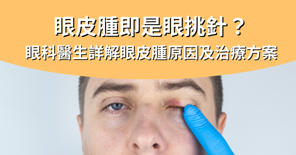
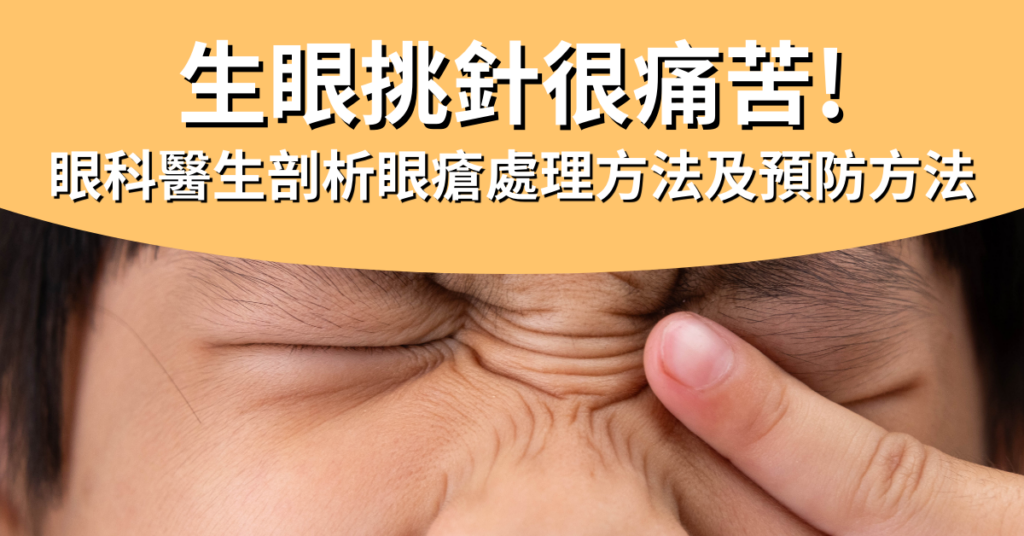
Picking needles in raw eyes is painful! How many patience first? Ophthalmologist analyzes how to treat eye sores and how to prevent them

Peeping into someone else's bath will cause eye sores/eye needles? How long do symptoms take to get better? Is it possible to recover by applying ointment on your own? Or do you want to see an ophthalmologist for eye sores/eye needles? I believe that many people will have similar questions, especially the most want to know, once sick, how long will eye sores / eye needles be good? Before answering these questions, we need to understand the pathogenesis.

Causes of eye sores/eye needles

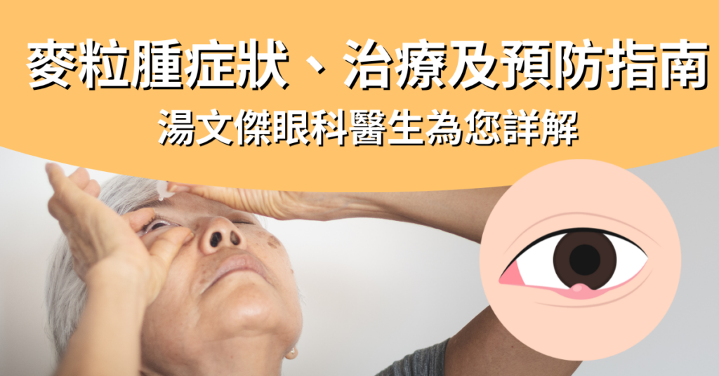
Eye sores, commonly known as eye needles, are medically called "styes". Eye sores/needles usually occur near the meibomian glands and can cause redness and swelling of the eyelids. The main cause of eye sores/needles is excessive oil secretion of the meibomian glands, resulting in blockage of the glands, coupled with the lack of attention to eye hygiene on weekdays, which causes bacterial infections.

In addition, if the patient often develops eye sores/needles, it may also be because the hair follicles of the eyelashes are blocked by mites. These tiny creatures can live at the roots of eyelashes, and when they grow in large quantities, they can lead to inflammation and the accumulation of pus, eventually forming eye sores/eye picks. So in fact, it is not formed because of peeking at other people's baths.

To prevent eye sores/needles, daily eye care and cleansing are very important, especially for people who wear makeup frequently, be sure to remove makeup thoroughly and avoid sharing makeup and makeup tools with other people as much as possible. If eye sores/needles are almost intolerant and tend to occur, an ophthalmologist should be consulted to ensure that the sores/needles condition is managed appropriately.

Characteristics and symptoms of eye sores/needles

Eye sores/needles grow in different locations and correspond to different names, namely "external styes" and "internal styes". The sebaceous glands that grow on the more superficial eyelashes are external styes; The internal styes grow in the deep meibomian gland, because the pus point is on the inside of the eyelid, so the inflammation time will be longer.

At the beginning of the disease, there is only a slight foreign body sensation, and it will not be painful to the touch, and such symptoms will heal themselves as long as they rest more and apply appropriate heat. However, if there is a bacterial infection with eye sores/needles, the following symptoms will occur:

1. Symptoms of eye sores/eye needles

  • Slight redness and swelling of the eyelids
  • Minor itching and dandruff
  • There is a foreign body sensation
  • A scratching sensation when blinking

2. Severe symptoms of eye sores/eye needles

  • The eyelids are visibly red, swollen, enlarged, and even affect the field of vision
  • Pain, eyelids accompanied by slight warmth
  • The foreign body sensation is heavy
  • Transient astigmatism is present

If persistent swelling and pain are found, an ophthalmologist should be consulted immediately for eye sores/needles.

How are eye sores/needles treated?

Some patients try to pluck the affected area with a needle and squeeze the pus out when dealing with eye sores/needles, but this may cause bacterial infection, causing the wound to become inflamed or recur again, or causing cellulitis or even brain infection. Therefore, doctors do not encourage patients with eye sores/needles to be treated in this way. If you find that you have eye sores/needles, you should consult an ophthalmologist. Usually, when doctors treat and treat eye sores/needles, they will provide appropriate treatment recommendations according to the severity of the patient.

1. Mild eye sores/eye needles

To deal with mild eye sores/eye pick-and-needles, as long as you pay attention to eye hygiene, you can try to warm towels and "hot eggs" to warm the compress to make the eye sores/eye pick-needles subside on their own, the method is to boil the eggs, do not need to peel the shell, wrap them in a gauze towel, gently massage the eyes, apply 4 to 5 times a day, each time for 15 minutes, the blocked oil will melt and push out, but also reduce the blockage of the hair follicles by the oil, this method of eye sores / eye picks needles will be good without a few resistances.

However, if you find that the eye sores/needles are too painful, it is recommended to seek help from a doctor who will also prescribe some steroids or antibiotics according to the eye sores to help reduce inflammation and prevent infection.

2. Severe eye sores/eye needles

If you notice that the eye sores/needle are still inflamed and do not improve, the doctor will recommend that the person with eye sores/needle pick needle inject corticosteroids to remove the lump. However, this medication whitens the surrounding skin and is not optimal for dark-skinned people, so doctors may recommend minor surgery to remove eye sores/needles. This type of surgery is fairly simple and the wound is minimal, and there is no scarring on the appearance. If eye sores/needles still occur frequently in the same area after surgery, doctors will send the tissue to a laboratory to rule out cancer, but usually lumps on the eyelids are benign and harmless.

How good is eye sore/eye needle?

As for eye sores/eye needles, it depends on the severity of the patient. Generally speaking, mild eye sores/eye needles should be patient and wait a few days to get better, and the swelling will slowly subside; Severe patients need to be more patient, and eye sores will not improve until almost 2 weeks. If eye sores/needles are treated with resistance, they will get better in almost a few days and the course of the disease is expected to be shortened.

Whether it is a mild or severe patient, when dealing with eye sores/eye needles, the most important thing is to seek medical attention in time, and be patient to wait a few days to see if it will improve, self-treatment of eye sores/eye needles is not recommended, because this will cause more problems, resulting in the final eye sores/eye needles are almost more patient first will improve, recover, then may even take more than a whole month to recover.

Are eye sores/needles contagious?

In general, eye sores/needles are not contagious by themselves. However, if you do not treat eye sores/needles in time, which can lead to the growth of bacteria or hair follicle mites, there is a risk that it may be transmitted to the other eye or to others through contact. Sharing towels, pillows, or cosmetics with people increases the risk.

Will eye sores/eye needles be almost intolerant and often recur?

Anyone who has long sores or needles knows that the feeling is not only quite uncomfortable, but sometimes directly affects the appearance. Although this is not an emergency, it is definitely the biggest problem for some people, because eye sores/eye needles are almost intolerant and often recur. To prevent and prevent recurrence of crises, the most important thing is to start with hygiene, diet and preventive treatment.

  • Pay attention to hygiene: keep your hands clean, especially before touching your eyes; Also avoid touching your eyes with your hands and wiping your eyes with unclean towels
  • Healthy work and diet: reduce staying up late and let the eyes fully rest, in order to effectively regulate the oil secretion of the meibomian glands; Reduce the consumption of fried or spicy foods to reduce the chance of inflammation in the body

TREATMENT TO PREVENT RECURRENCE IS MORE EFFECTIVE IN PATIENTS WHO HAVE RELAPSES THAT ARE ALMOST INTOLERANT TO EYE SORES/NEEDLES, AS THEY KILL PARASITES, ARE ANTI-INFLAMMATORY, AND ANTIMICROBIAL. TAKING IPL (INTENSE PULSED LIGHT) AS AN EXAMPLE, IPL HAS PHOTOBIOREGULATORY FUNCTIONS, EFFECTIVELY STIMULATES CELL MITOCHONDRIA AND PROMOTES CELL HEALTH. TREATMENT WITH ANTIBIOTICS CONTAINING TETRACYCLINE OR MACROLIDES CAN HELP WITH ANTI-INFLAMMATORY AND BACTERIOSTATIC TREATMENT.

If not properly managed, eye sores/needles may be tolerant and often recur after a few months, or cause other more serious eye problems. Therefore, if there is eye sore/needle picking, it is best to seek an ophthalmologist to manage eye sores/eye needles.

Ophthalmologists analyze the treatment methods and prevention methods of eye sores Read More »

Featured Image_Will age-related macular degeneration get better?

Will age-related macular degeneration be okay?| Ophthalmologist says you know: senile macular disease becomes the cause and treatment method

Macular degeneration is a chronic eye disease that is often referred to as age-related macular degeneration due to blurred central vision, distorted images, and dark spots in the central area, mainly caused by the gradual aging of the eyes with age. However, some young people may develop age-related macular degeneration due to other factors. So, with current medical technology, is there a feasible solution to treat age-related macular degeneration? Will some macular degeneration be better on their own? The following is to explain the causes of age-related macular disease and surgical treatment, as well as early detection of eye problems, and early surgical treatment of age-related macular degeneration.

Type of macular degeneration

The macula is located in the middle of the retina, and most of the photoreceptor cells are concentrated in the macula, which can help us clearly see things in front of us, such as words, colors, and appearance. Clinically, macular degeneration is divided into two morphologies: dry age-related macular degeneration and wet age-related macular degeneration, of which wet age-related macular degeneration is more severe than dry age-related macular degeneration, and the treatment is also different.

Dry age-related macular degeneration

  • Dry age-related macular degeneration is the most common type, accounting for about 8~9%.
  • The cause is the accumulation of metabolites formed by the degeneration of retinal cells
  • Dry age-related macular degeneration progresses slowly and may not have a noticeable visual impact at first, but as the disease progresses, vision may gradually become blurred or lose central vision, and even develop into wet age-related macular degeneration

Wet age-related macular degeneration

  • Age-related macular degeneration is less common, and people with these macular degeneration may become blind
  • The cause is the growth of abnormal blood vessels in the macular area, resulting in the destruction of all layers of the retina, resulting in retinal hemorrhage, vitreous hemorrhage, macular edema, and retinal scarring pull
  • Age-related macular degeneration progresses more rapidly and may cause sudden blindness or severe vision loss in a short period of time

What causes age-related macular degeneration?

Senile macular degeneration is affected by a variety of factors, the most important of which is age-related, which is the main cause of central vision loss in middle-aged and elderly people over 50 years old. However, some young people may also develop macular degeneration, which may be due to congenital inheritance or acquired bad habits. Other factors include:

  • Smoking: Studies have shown that smokers are 3 to 4 times more likely to develop macular degeneration than the general population
  • Poor eating habits: Long-term intake of high-fat, high-sugar and other foods causes obesity and three highs, affects the health of eye blood vessels, and increases the risk of macular degeneration
  • LONG-TERM EXPOSURE TO INTENSE SUNLIGHT: UVA IN THE ULTRAVIOLET RAYS OF SUNLIGHT ACCELERATES THE AGING OF PHOTORECEPTOR CELLS, CAUSING DAMAGE TO THE MACULA
  • CELL GROWTH FACTOR ROLE: SOME GENETIC ABNORMALITIES IN THE HUMAN BODY OR RELATED TO MACULAR DEGENERATION, SUCH AS COMPLEMENT FACTOR B, COMPLEMENT FACTOR H, THESE GENE ABNORMALITIES HAVE BEEN SHOWN TO BE GREATLY RELATED TO THE DEVELOPMENT OF MACULAR DEGENERATION

In addition, although blue light from electronic products is rumored to be one of the causes of age-related macular degeneration, there is not enough research data to confirm this claim. However, using electronics in a darker environment may cause the macula to absorb more blue light, which can lead to various eye diseases.

What are the symptoms of age-related macular degeneration?

The symptoms of age-related macular degeneration may only occur in a single eye, but it is also possible that both eyes appear successively, if you find that the central vision is a little blurry, the image is distorted or the color becomes lighter, etc., you can basically determine that it is age-related macular degeneration, but it is still recommended to go to an eye clinic for examination to make a correct judgment.

  • Visual disturbances: Age-related macular degeneration affects central vision, and people feel that central vision gradually becomes blurred or has dark spots, making it difficult to recognize details, read, drive, and distinguish faces
  • Altered visual field: patients may feel things deformed in the central field of view, for example, straight lines may appear curved or distorted; Distance and altitude are often misjudged
  • Abnormal color vision: People with age-related macular degeneration may have problems with color perception, such as dull colors or difficulty distinguishing similar colors

MACULAR DEGENERATION SELF-TEST METHOD - AMSLER GRID

AMSLER GRID IS A QUICK WAY TO DETECT AGE-RELATED MACULAR DEGENERATION AT HOME, AND IF YOU FEEL YOU ARE AT RISK OF AGE-RELATED MACULAR DEGENERATION, YOU CAN FOLLOW THESE STEPS:

  1. Find a bright place with your eyes about 30cm away from the checkered table
  2. Cover one of the eyes with your hand and stare at the black dot in the center
  3. Repeat the above action with the other eye

If you notice that the straight line is curved, some areas are blurred or missing, or it may be a warning sign of age-related macular degeneration, you should seek advice from your ophthalmologist as soon as possible.

However, although age-related macular degeneration causes a decrease in central vision, it does not affect peripheral vision, and the risk of complete blindness is relatively low. Although age-related macular degeneration does not cause complete blindness, loss of central vision can have a considerable impact on activities of daily living. Once age-related macular degeneration is suspected, you should seek immediate consultation with an ophthalmologist for early treatment to control the condition of age-related macular degeneration.

What are the surgical treatments for age-related macular degeneration?

There are laser surgery and intraocular injections for the treatment of age-related macular degeneration, and the cost of surgery for both types of macular degeneration is also very valuable. At present, intraocular injection is the most mainstream method of treating age-related macular degeneration, and for some more serious patients, doctors will recommend surgery to treat age-related macular degeneration.

In addition, most of the treatment options for age-related macular degeneration focus on more severe wet age-related macular degeneration, and most doctors will recommend that patients consume more eye-friendly foods or supplements on a daily basis, change bad habits such as smoking, and get regular check-ups.

Laser light coagulation

In addition to the treatment of age-related macular degeneration, laser photocoagulation surgery can also treat other eye diseases such as cataracts, and its principle is to use the heat energy of the laser to destroy the abnormal blood vessels, so as to reduce blood vessel bleeding and prevent vision loss. However, this age-related macular degeneration treatment may destroy healthy tissue, and if the blood vessels are located fairly close to the center of the macula, it is advisable to find another treatment option.

Photodynamic therapy

Photodynamic therapy is a combination of drugs and lasers to treat age-related macular degeneration, in which the ophthalmologist will first inject photosensitive drops into the patient's arm, which will follow the body's blood flow and attach to the abnormally proliferated blood vessels, and then use cold lasers to stimulate the photosensitive drops, which will destroy the abnormal blood vessels, thereby achieving the effect of treating age-related macular degeneration. Unlike laser photocoagulation, this type of macular degeneration treatment surgery does not harm the surrounding tissues and is also beneficial for improving vision problems.

Anti-vascular endothelial growth factor vitreous injection

Injections in the eye are the most commonly used methods for the treatment of age-related macular degeneration, the ophthalmologist will administer an anesthetic injection on the surface of the eyeball, and then inject anti-vascular endothelial growth factor to prevent abnormal vascular hyperplasia, thereby reducing the risk of neovascular bleeding, this kind of injection treatment can not only control the condition of macular degeneration, but also improve the damaged vision, injection is quite effective in the treatment of wet age-related macular degeneration.

Another intraocular injection for age-related macular degeneration is steroid injection, which can also effectively reduce the damage of the retinal barrier by intraocular blood vessels and improve macular edema. However, it is important to note that steroids have the potential to cause other eye problems such as increased eye pressure, cataracts, etc.

New technology for the treatment of advanced age-related macular degeneration

When age-related macular degeneration cannot be treated with lasers or injections, ophthalmologists often recommend some assistive methods to improve vision, such as bifocal additional intraocular lenses in the eye or low vision glasses. Although this method may be more expensive than macular surgery, it is relatively straightforward and quick.

Low vision glasses

Smart low vision glasses project images of their surroundings in real time to the healthiest areas of the retina of low vision people and adjust them to the patient's perception. These glasses improve the patient's perception of real-world scenes by supplementing and correcting visual details. During the setup process, the ophthalmologist can intuitively adjust the glasses to help the patient find the best vision.

Bifocal additional intraocular lens

SML (Scharioth Macular Lens) is an implantable lens that can help patients "magnify" their field of view, with a specially designed central optical area that provides a high additional optical focal point of +10.0D to improve advanced vision without compromising distance vision.

Will some macular degeneration get better after surgery?

Some patients ask, "Will macular degeneration be better after surgery?" Or "Will the macular degeneration get better on its own?"

Unfortunately, age-related macular degeneration is an eye disease that cannot heal naturally, and surgery or other age-related macular degeneration treatment options can only slightly improve vision and prevent it from deteriorating further. The key is prevention, usually should take care of eye health, reduce direct eyes, supplement more nutrients such as lutein, try to delay the speed of eye aging, in order to reduce the chance of suffering from age-related macular degeneration.

After completing any macular surgery, be sure to follow your doctor's instructions to order or take your medication on time. But is it possible that as long as you take medicine on time, the condition of macular degeneration will be completely fine? In fact, patients should also pay more attention to their daily life, try to avoid rubbing their eyes, reduce eye use, and avoid smoking or construction sites and other soot-heavy environments. Macular degeneration surgery should also pay attention to diet and consume more nutrients that are beneficial to the macula, such as dark green vegetables, goji berries, egg yolks, etc. Studies have shown that daily intake of 6 mg of lutein effectively reduces the chance of macular deterioration. If the patient suffers from age-related macular degeneration due to three highs or other health problems, it is necessary to change the diet and lifestyle to control the recurrence of macular degeneration and reduce the risk of recurrence.

Will age-related macular degeneration be better? | What do ophthalmologists say: age-related macular disease becomes the cause and treatment Read More »

Explain Presbymax presbyopia laser vision correction surgery

Get rid of reading glasses from now on! 【Presbymax Presbymax Laser Surgery】Helps restore youthful vision

Presbyopia is a vision problem that occurs with age, and almost everyone has presbyopia, usually in people around the age of 40. Many people with presbyopia are reluctant to wear reading glasses for fear of being ridiculed for their age, which affects their daily lives. However, now that there are more and more ways to correct presbyopia, wearing reading glasses is not the only option, such as the Presbymax presbymax laser surgery highly recommended by ophthalmologist Dr. Tang Wenjie, which can not only avoid the embarrassment of wearing reading glasses, but also restore the vision of the eyes when they were young.

What is presbyopia?

The most common symptom of presbyopia is dyslexia, reading the text on the mobile phone or newspaper can become quite strenuous, and it is necessary to constantly adjust the distance to find a clearer focus. Prolonged close movement can make it easy to feel eye strain, dryness or discomfort; When working at close range in darker environments, more light is needed to help the eye focus.

The main cause of presbyopia is the degeneration of the lens and ciliary muscle in the eyeball. This degradation causes the lens to become stiff and inelastic, which in turn affects the eye's refractive ability and ultimately vision.

Although presbyopia usually occurs in people in their 40s, however, with the advancement of technology, people are increasingly relying on electronics. Staring at the screen for a long time will cause the ciliary muscle to tighten too much, causing the eyes to enter the presbyopia stage early. Therefore, protecting eye health is extremely important for modern people. It is recommended to reduce the time spent using electronic products to allow enough time for the eyes to relax to slow down the development of presbyopia.

2. Method self-test for presbyopia

Method 1: Symptom check

The following are some common symptoms related to presbyopia, if you get more than 3 of them, you may suffer from presbyopia.

  • When looking at the phone, you need to hold farther away to see clearly
  • When looking at the subject, I want to take off my glasses, but I can see more clearly without glasses
  • Feel that your home or office is darker than before
  • Often misread numbers or words
  • Eye strain is often felt
  • Shoulder and neck aches, stiffness
  • Often frowning, dizziness

Method 2: Banknote test

  1. Take out a note and place it where you can see it best
  2. Then focus on the small print on the banknote
  3. Look away from the banknote and look at an object 2 to 3 meters away for about 5 seconds
  4. Then look back at the small print and see if you can read the small print in a few seconds

Being able to focus within 1 second is normal; More than 1 second is at risk of presbyopia; More than 3 seconds can basically determine that there is presbyopia.

If you suspect that you have presbyopia, it is recommended to consult an ophthalmologist as soon as possible for professional advice and help. Your ophthalmologist will give you the need to wear reading glasses based on your vision needs or recommend Presbymax laser surgery to improve presbyopia. Early recognition and treatment of presbyopia can help improve vision and improve quality of life.

What is the difference between presbyopia and farsightedness?

Presbyopia and farsightedness are also blind, but the causes of the two are not the same. In addition, if you find that you have vision problems, it is recommended to go for a test.

  • Presbyopia is the degeneration of the eyeball

Presbyopia is an age-related vision problem in which the lens and ciliary muscle of the eye gradually deteriorate with age, making it less focused and making it difficult for the eye to see clearly in close objects and dark environments. If presbyopia has seriously affected your life, Presbymax presbyopia correction surgery can be considered. This is because Presbymax can not only treat presbyopia, but also help improve myopia, farsightedness, astigmatism and other problems, and restore the eyes to their youthful state.

  • Farsightedness is innate

Farsightedness is a vision problem related to the structure of the eyeball. It is mainly formed by short eyeballs or weakness of the ciliary muscle. If only the eyeball is short but the ciliary muscle is developed, there will basically be no problem of blurred vision; When the ciliary muscle is weak, it needs to be forced to focus on the near object. Farsightedness is innate, meaning that most children have farsightedness, but there is no need to worry too much, as the eye develops normally, the farsightedness will be relieved.

Leave the old flowers alone, be careful of physical problems!

Many patients feel that presbyopia has little impact on life and are reluctant to face up to the problem of presbyopia, thinking that they can see clearly by simply squinting, and even reading glasses are reluctant to wear. However, this behavior will cause ciliary muscle overwork, increase the burden on the eyes, cause eye soreness and discomfort, eye pain, etc., and in severe cases, it will even spread to the shoulder and neck area, resulting in shoulder and neck pain, headache, dizziness, nausea and other symptoms.

Although presbyopia is not an emergency, the degree of presbyopia will increase with age. If presbyopia at 100 degrees is diagnosed at 40 degrees, it will increase to 300 degrees at 60 degrees, and the degree of presbyopia will increase by about 10 degrees per year. If the presbyopia degree decreases abnormally, this may be a warning sign for cataracts, and patients should seek detailed examination and appropriate correction from an ophthalmologist as soon as possible. Timely identification and treatment of presbyopia is the key to protecting eye health.

What are the ways to correct presbyopia?

With the continuous advancement of medicine, more and more ways to correct presbyopia, including monofocal glasses, multifocal lenses, contact lenses or surgical implantation of intraocular lenses, Presbymax presbyopia laser surgery, etc.

  • Monofocal glasses: Usually monofocal glasses only need to be worn when looking at things up close. Such glasses can only provide clear vision at a specific distance. However, if a person has both myopia and presbyopia, then it is necessary to frequently remove and wear two glasses of different degrees in different situations, and then multifocal glasses can be considered.
  • Multifocal glasses: These glasses are equipped with multiple lenses with different focal points, allowing you to see far, medium and close distances in one pair of glasses without the need to change glasses frequently.
  • Multifocal contact lenses: If you don't like the bulkiness of glasses or people who are used to wearing contact lenses, you can use multifocal contact lenses or directly have surgery to correct your vision.
  • Surgical treatment: surgical treatment can include monocular vision surgery, multifocal intraocular lens surgery, Presbymax presbyopia laser correction surgery, etc. Presbymax presbyopia laser correction surgery is a relatively new option. This procedure uses laser technology to reshape the cornea to improve presbyopia vision, thereby reducing or eliminating dependence on glasses or contact lenses.

Traditional vision correction surgery VS Presbymax presbyopia correction, which one to choose?

Monocular vision surgeryMultifocal intraocular lensPresbymax presbyopia
❌ Medium-range images are blurry
❌ Poor three-dimensionality❌ There is a glare problem
❌ Not suitable for night motorists
❌ It takes time to get used to✅ Far, medium and close vision is clear
✅ No glare issues
✅ No time is required to adapt, and normal activities can be carried out 24 hours after surgery

Presbymax presbyopia surgery mainly uses laser polishing on the surface of the cornea to make the cornea multifocal state. The main operation is to correct the myopia, farsightedness, astigmatism, etc. of the main eye to close to 0 degrees; The other eye retains or creates myopia of about 150 to 250 degrees, which is dedicated to seeing close distances and improving presbyopia. In addition, Presbymax also adds Spherical Aberration to increase the depth of field of both eyes, extend focus, and change chromatic aberration, thereby creating a 'fusion zone' for both eyes, allowing patients to obtain clear vision at the same time, and ensure the original color and stereoscopic acuity.

Dr. Tong said that Presbymax is one of the most recognized and mature presbyopia correction methods in the world. Patients who have undergone Presbymax presbyopia surgery are extremely satisfied and the price is relatively affordable. The procedure is very short, about 10 minutes to complete, within 24 hours the patient can move normally, within a few weeks vision can be fully restored, no discomfort, so Presbymax can be the best choice for presbyopia.

Get rid of reading glasses and Presbymax to help restore youthful vision Read More »

Is a black spot in the eye a precursor to blindness? Ophthalmologist explains in detail the treatment and improvement of floaters

Floaters are sometimes annoying, people with floaters will see "black spots" floating in their eyes, not painful or itchy and unable to rub open, when you look directly at them, they will disappear in an instant. Floaters come in different shapes, such as black dots, lines, circles, ovals, tadpoles, etc., especially when looking at a bright and clean background, it is easier to spot its presence. Floaters are a degenerative disease of the eyes, mild patients in general, as long as they are used to coexisting with black spots and floating objects in the eyes, daily life is not affected, they do not need to receive floaters treatment, but if the black spots or floating objects in the eyes are too large or too much, floaters suddenly attack or flash, so that vision is damaged, you should seek improvement methods, find an ophthalmologist for the treatment of floaters for consultation and surgery or laser treatment of floaters, otherwise, the longer it drags on, the more serious or blind!

What is floaters?

Floaters are medically a phenomenon of vitreous degeneration of the eye. The vitreous is a transparent gel located behind the lens and before the retina. Normally, the vitreous fills the entire vitreous cavity to support the shape of the eyeball. But with age, myopia and other problems, the vitreous will liquefy and shrink, forming turbid fibers, and these fibers will float around in the vitreous, when the light refracts these fibers, the patient will feel that there are black spots in the eyes, which is the precursor of floaters.

Illustrated floater lesions

Floaters are medically a phenomenon of vitreous degeneration of the eye. The vitreous is a transparent gel located behind the lens and before the retina. Normally, the vitreous fills the entire vitreous cavity to support the shape of the eyeball. But with age, myopia and other problems, the vitreous will liquefy and shrink, forming turbid fibers, and these fibers will float around in the vitreous, when the light refracts these fibers, the patient will feel that there are black spots in the eyes, which is the precursor of floaters.

Who is most likely to develop floaters?

Floaters usually become more common with age, so older people are more likely to have dark spots in their eyes. But in addition to age, other factors that may increase the risk of floaters include myopia, history of eye surgery, eye trauma or eye inflammation. According to clinical studies, high-risk groups for floaters include:

  • Middle-aged and elderly
  • People with high myopia
  • People with hypertension/diabetes
  • Have had eye surgery
  • Blows to the head, e.g. car accidents, divers
  • Other eye problems, such as eye inflammation

However, most floaters are benign, such as because of old vitreous degeneration, or excessive eye fatigue, there may be symptoms of floaters, as long as there are more black spots in the eyes, and the position is fixed, do not worry too much.

Conversely, if suddenly there are a large number of dark spots in the eye that have affected the field of vision or there is a flash, it may be vitreous peeling, resulting in pulling the retina to the hole or even falling off. This condition must be investigated immediately, for improvement, or for floaters.

Causes of floaters

The causes of floaters can be broadly divided into three types, namely physiological, degenerative and pathological.

  • Physiological

About 80% of floaters are physiological. Physiological floaters usually occur in people under the age of 40 or people who have been using their eyes for a long time, and most people will see black spots in the eyes, which are vitreous impurities that usually do not affect vision, and do not need to be treated immediately for floaters, and will disappear on their own over time

  • Degenerative

As we age, the vitreous body of the eyeball, like other organs in the body, gradually deteriorates. In this degeneration process, the vitreous body will shrink to form fine fibers, these fibers will float in the glass, when the light enters the eye, and these impurities refract, it is the black spots in our eyes.

  • Pathological

Pathological floaters are caused by eye disease or systemic vascular disease. The so-called eye disease refers to the retina under the pull or degeneration of the process of the retina in the process of tearing or degeneration, resulting in vitreous hemorrhage, and even retinal peeling, resulting in visual impairment, and in the worst case, permanent blindness. In addition, systemic vascular diseases, such as eye stroke, diabetes, hypertension or macular degeneration, if not paid attention to and actively treated, may also appear pathological floaters, patients should seek to improve floaters as soon as possible.

Complications of floaters – retinal detachment/tear

Although suffering from floaters is not a serious disease, retinal detachment caused by floaters is an emergency in ophthalmology. One in four people with floaters may have their vision affected by retinal detachment or a tear. Keep in mind that if you have these signs of retinal detachment due to floaters, including:

  • There are a large number of dark spots in the eyes in a short time
  • Unusual flashes
  • shadow
  • The view is blocked

This means that there may be cracks in the retina, and it is important to seek immediate medical attention for treatment, otherwise floaters will cause permanent damage to vision or even blindness.

How is floaters treated?

If you have benign floaters, you do not need to be treated immediately. When you notice dark spots in your eyes, you can try moving the eyes to let the fluid flow through the eyes so that the fibers leave the line of sight. Of course, some people can't bear the black spots floating around in their eyes, and their attention is often disturbed by black spots, which seriously affects their mood, so they can consider floaters treatment.

With current technology, the main treatment and improvement of floaters is laser therapy and vitrectomy.

First, laser treatment of floaters

This floater treatment is suitable for large and concentrated vitreous fibers, using laser light to break these fibers into smaller fragments, through these methods to improve and eliminate floater symptoms. The laser treatment is performed under local anesthesia without pain and the whole treatment takes only 15 to 20 minutes. However, not everyone is suitable for laser treatment to improve floaters, and if the fibers are loose or too close to the macula or crystals, it should not be performed to avoid complications such as cataracts or macular damage.

Second, vitrectomy surgery to treat floaters

Vitrectomy is performed by removing the vitreous inside the eye through a small incision and replacing it with a solution to maintain the shape of the eye. The procedure only takes about 10 to 15 minutes. However, vitrectomy does not necessarily completely remove the fibers, and if the surgery itself causes bleeding or retinal tears, new floats may form, so most physicians do not recommend this procedure to treat floaters.

Floaters improvement and maintenance methods

Once floaters occur, it is almost difficult to reverse, even after laser or surgical treatment, there is a possibility of recurrence, what we can do is to live peacefully with it, or we can improve floaters by the following methods to reduce symptoms slightly.

First, the daily maintenance of benign floaters

  • Avoid excessive eye use

PAY ATTENTION NOT TO OVERWORK THE EYES, ESPECIALLY 3C HEAVY USERS, YOU SHOULD LET THE EYES REST FOR 5 TO 10 MINUTES EVERY 1 HOUR, USE THE REST TIME TO BLINK, LOOK INTO THE DISTANCE TO RELAX THE EYES.

  • Good living habits

In daily life, it is recommended to reduce the time spent playing with mobile phones, especially to avoid staying up late at night to look at mobile phones, to ensure that you have enough sleep and do exercise in a timely manner, which is also a way to improve floaters.

  • Regular follow-up

Do not think that you have benign floaters can relax, floaters must be regularly checked to avoid other lesions, miss the best time for treatment.

  • Eye care diet

Foods with antioxidant effects, such as berries, green or yellow vegetables, carrots, soybeans, milk, and fish oil, should be consumed in your diet to help improve floaters.

2. Postoperative care and maintenance of malignant floaters

  • Medication treatment

After surgery, be sure to follow the doctor's instructions, remember to wash your hands thoroughly with soap before each medicine to avoid cross-infection.

  • Improve your lifestyle

Rapid eye movements and excessive eye use should be avoided for 1 week after surgery. Try not to read books, mobile phones, computers, TV, etc. during this period. In addition, it is recommended not to do strenuous exercise, climbing, diving, or flying, as these activities can irritate the eyes and affect the speed of postoperative repair.

  • Use an eye patch

During eye repair, it is recommended to wear an eye mask at all times to prevent collisions and avoid unconsciously rubbing your eyes when sleeping at night. It is necessary to ensure that the eye mask is clean when using, and it is recommended to wash and disinfect the eye mask daily to avoid wound infection due to bacteria.

  • Daily diet

In addition to avoiding irritating foods, it is also necessary to avoid eating legumes, because such foods are easy to let the nitric oxide in the blood run into the eyes, causing the inert gas in the eyes to produce flatulence and increase the pressure in the eyes.

  • Safe at home

Keep your home clean and tidy, reduce dust and avoid secondhand smoke, and do not change the layout of the home or place objects in the aisle during the recovery period, so as not to cause collisions or falls in unfamiliar environments and irritate the eyes.

  • Regular follow-up visits

In addition to following the doctor's advice for regular follow-up consultations after surgery, if you notice eye discomfort, such as persistent eye pain, sudden blurred vision, nausea, increased or larger dark shadows, etc., remember to consult your ophthalmologist immediately to avoid worsening of the condition.

How can floaters be prevented?

Although we cannot completely avoid the occurrence of floaters, we can slow down the aging of the eyes and reduce the chance of developing floaters through daily maintenance.

1. Develop good eye habits

  • AVOID EXCESSIVE USE OF 3C PRODUCTS (1 HOUR WITH EYES AND 5-10 MINUTES BREAK)
  • Works in sufficient light and stable without flickering
  • Avoid staying up late to increase the burden on your eyes

2. Intake of eye care nutrients

  • Carotenoids (dark green, dark yellow and red fruits and vegetables)
  • Anthocyanins (plum fruits)
  • VITAMINS (VITAMINS A, B, C AND E)
  • Omega3 fatty acids (fish oil)

3. Proper control of systemic vascular diseases (such as: diabetes, hypertension)

  • Maintain a balanced nutrition and reduce the intake of foods high in oil and salt and sugar
  • Regular exercise (jogging, cycling)
  • Have regular full-body examinations

Although most floaters can not be treated, they should not be completely ignored, and they must be checked regularly, and if symptoms are aggravated, they should seek medical attention immediately to avoid missing the golden period of floater treatment.

Is a dark spot in the eye a sign of blindness? Read More »

Cover image_Comparison_Glaucoma_Macula_Cataract

【TREATMENT OF EYE DISEASES】GLAUCOMA VS MACULAR DISEASE VS CATARACT OPHTHALMOLOGISTS TEACH EYE DISEASE SYMPTOMS + TREATMENT METHODS

Elderly people often have eye blindness, and may think that it is a degenerative problem that makes it difficult to see clearly, but have they ever thought that they may suffer from eye diseases due to eye lesions?

Eye disease symptoms

First of all, to distinguish whether vision suddenly or slowly decreased, visual impact is the middle range, the whole picture, or peripheral vision? And is there a feeling of pain in the eyes? So how to tell the difference between macular disease, cataracts and glaucoma? Taking cataracts as an example, the vision of the average elderly will slowly decline, and it will affect the whole picture. Macular disease, on the other hand, has a sudden and acute decrease in vision, affecting central vision. Glaucoma also slowly affects vision, generally affecting peripheral vision first.

These three diseases can be easily confused, and there are many ways for the elderly to check them. There is a kind of "checkered paper". Checkered paper is a kind of checkered paper, and the elderly can see whether the straight line has changed the curve by looking at the checkered paper to see if the straight line has changed the curve, and these conditions can determine whether there is macular disease. For other problems, an eye disease can only be determined through an ophthalmologist's examination.

How to heal?

There are generally two complications of the upper eye of diabetes, the main cause is hypoxia, resulting in angiogenesis. If angiogenesis occurs, those fragile blood vessels may have a condition that leaks, leading to vitreous hemorrhage or macular edema. To prevent this, it is usually necessary to inject eye drugs to prevent angiogenesis, thereby reducing hypoxia.

Cataracts are basically performed through minimally invasive cataract surgery, in which cloudy lenses are removed and implanted with intraocular lenses. Finally, glaucoma, with early treatment, the nerves can be controlled so that they do not continue to be damaged. There is no treatment for glaucoma, only testing, medication, surgery or laser to control intraocular pressure to slow down the damage to the nerves.

ELDERLY PEOPLE CAN PREVENT IT BY CHANGING THEIR DAILY LIFESTYLE, SUCH AS: LESS SUGAR AND LESS SALT; EAT FOODS RICH IN VITAMIN A, CAROTENE OR OMEGA FATTY ACIDS, BECAUSE OF ANTIOXIDANT EFFECTS; DO AT LEAST 30 MINUTES OF AEROBIC EXERCISE; DO NOT SMOKE. THE ELDERLY NEED REGULAR EYE EXAMINATIONS FOR EARLY PREVENTION AND EARLY TREATMENT. TRY TO AVOID ULTRAVIOLET RAYS TO IRRADIATE THE EYES TO PREVENT VARIOUS EYE DISEASES.

Source: Topic

【Treatment of eye diseases】Glaucoma VS macular disease VS cataract Ophthalmologists teach eye disease symptoms + treatment methods Read More »

Cover image_Red eye pain_Hidden disease_Rheumatoid arthritis_Eye inflammation

【Eye inflammation】Red and painful eyes or reflect hidden diseases in the body Doctor: 80% of rheumatoid arthritis patients have eye inflammation

Many doctors will use the eyes as a window to observe physical diseases, especially long-term patients, which diseases will affect the eyes?

Diseases that affect the eyes

Eye pain, redness, eye inflammation and other conditions, ordinary elderly people may feel, but if the situation continues, we will worry about other hidden diseases in the body, one of which is an autoimmune system disease, which will cause various inflammations in the body. The eye is also one of the affected organs, and if not looked at or treated, the eye's vision will continue to decline and there is even a risk of blindness.

Symptoms and treatment of immune system disorders

Immune system disorders occur in people of all ages, most commonly rheumatoid arthritis, xexia, ankylosing spondylitis, or lupus erythematosus. Taking rheumatoid arthritis as an example, patients may have low back pain, hip pain or inflammation, so that they have a sore back and a stalk. Calcification occurs due to inflammation. Joints will combine with each other, so that it is difficult to get up in the morning or want to bend over and reach for the toes, which will affect joint movement for a long time. First-line treatment is systemic and is usually prescribed by a rheumatoid specialist with oral anti-inflammatory drugs.

Steroids are generally used in first-line treatment, but if we use large doses of steroids, or if we continue treatment, we will generally switch to second-line drugs to reduce the side effects caused by steroids. Biologics and immunosuppressants are used as second-line agents to control long-term therapy.

Studies have shown that eighty percent of patients have at least one chance of eye inflammation in their lifetime, iritis is one of the inflammations, it will release cells, resulting in vision loss, and these cells, will bind to the protein in the eyeball, accumulate in the anterior chamber angle, resulting in the anterior chamber angle water failure to flow away, so that intraocular pressure rises, damage nerves and form glaucoma.

TO BE TREATED, BLOOD IS USUALLY DRAWN TO FIND INFLAMMATORY FACTORS AND X-RAYS, SUCH AS LUNG X-RAYS OR LUMBAR BONE X-RAYS, TO SEE IF THERE ARE LUNG OR LUMBAR BONE PROBLEMS. IF PRESENT, A SYRUP IS USUALLY GIVEN TO CONTROL INFLAMMATION (AN ANTI-INFLAMMATORY STEROID EYE DROP). IN ADDITION, TO SEE IF THERE ARE SIDE EFFECTS AND COMPLICATIONS, USE GLAUCOMA EYE DROPS TO CONTROL INTRAOCULAR PRESSURE, SO OPHTHALMOLOGISTS AND RHEUMATOID SPECIALISTS WILL WORK TOGETHER TO CARE FOR AND TREAT PATIENTS.

lotion

There are different types of potions, some potions are ordinary moisturizing potions, which do not help the condition, and sometimes delay treatment, and some potions have the effect of constricting blood vessels, but it is not controlling inflammation, so there is a chance to mistakenly think that the eye condition continues to improve; Finally, if the treatment dose is insufficient or the medication is not used for enough time, it will delay the treatment and cause irreversible problems in the eyes.

Coronavirus

Conjunctivitis is accompanied by respiratory infections, such as influenza or viral colds, which is actually one of the symptoms of new coronary pneumonia. The most common symptom of conjunctivitis is conjunctival hyperemia or redness and inflammation of the corn of the eyeball. Studies have shown that 30 new crown patients had a test during admission and found that one of them had symptoms of conjunctivitis, so it was closely related.

To prevent the new crown pneumonia, first of all, we must wash our hands frequently, wash with water and lobe liquid for at least 20 seconds, in addition, we need to maintain a proper social distance of 1.5 meters, and we need to maintain air circulation to protect ourselves in the new crown pneumonia.

Source: Topic

【Eye inflammation】Red and painful eyes or reflect a hidden disease in the body Doctor: 8% of patients with rheumatoid arthritis have eye inflammation Read More »

Cover image_New coronavirus symptoms_Pink eye_Ophthalmologist Dr. Tong Man Kit

Is pink eye also a coronavirus symptom?

Is pink eye also a symptom of the new coronavirus?

In the face of the new crown epidemic, everyone focuses more on epidemic prevention, infection symptoms and rehabilitation treatment, and less attention is paid to the relationship between infection and vision risk. It turns out that pink eye is also an early sign of the new crown pneumonia virus, and people are worried about whether droplets will be infected by falling into the eyes, whether wearing glasses has an effect on epidemic prevention, and other mysteries, the following are answered by ophthalmologists one by one.

Mild new crown pneumonia patients, common symptoms include runny nose, headache, fatigue, throat discomfort, cough, and even muscle pain, vomiting, diarrhea and other symptoms, there is a finger pink eye (medical name conjunctivitis) is also an early sign of the new crown pneumonia virus, which is true? Ophthalmologist Dr. Tong Man Kit explains: "Pink eye or conjunctivitis accompanied by upper respiratory tract infections (colds, flu, etc.) may also be signs of new coronavirus infection. A common symptom of conjunctivitis is hyperemia or redness and inflammation of the conjunctiva of the eye, a study published in the Journal of Medical Virology studied 30 patients admitted to the hospital with the new coronavirus, and one of the patients was diagnosed with conjunctivitis."

Pink eye can cause different eye diseases

According to the above study, although the risk of conjunctivitis in patients is low, in addition to conjunctivitis, the new coronavirus has been reported to be related to other eye problems, including episcleritis, uveitis, inflammation of the lacrimal glands, retinal and optic nerve inflammation, although also rare, but some of these problems may lead to vision loss.

People wear masks when they go out every day, but they are most worried that droplets of the new coronavirus will fall into their eyes and cause infection, Dr Tong said, ocular transmission has not been confirmed by research, but the mucous membranes distributed in many body cavities and organs, including the respiratory tract, are usually the most susceptible to infection with the new coronavirus and viruses. "The surface and inner eyelid of the eye are also called the mucous membrane of the conjunctiva, so if infected droplets fall into the eye, they may be susceptible to infection." Published reports suggest that severe acute respiratory syndrome coronavirus type 2 (SARS-CoV-2) may be transmitted through aerosol-conjunctival contact.

Goggle protection is superior

Thinking that glasses and sunglasses could block the barrier, it turned out to be "a misunderstanding", Dr. Tong said: "Neither glasses nor sunglasses can completely block respiratory droplets sprayed in your direction, but there are goggles that protect the sides and around the eyes, which can provide better protection." In fact, the mode of transmission of the new coronavirus is still thought to be mainly through respiratory droplets between people, but the virus can also survive for days on the surface. Therefore, touching an infected surface and then touching your eyes, nose, or mouth without washing your hands can cause infection, but this risk is generally considered low.

So can cleaning your eyes with water or over-the-counter eye drops reduce the chance of infection? He pointed out that the surface of the eye has its own protective mechanisms, including antimicrobial proteins and natural lubrication, so daily cleaning or rinsing the eye with water is unnecessary and may actually wash away some of the natural protective barrier on the surface of the eye, increasing the risk of infection. If the eyes become inflamed or red, over-the-counter artificial tears may help relieve symptoms, but if symptoms persist, you should seek medical attention from your ophthalmologist as soon as possible.

Wear contact lenses and maintain hygiene

More than a myopic person has said that they are worried that wearing and removing contact lenses regularly increases the risk of contracting the new crown pneumonia virus, Dr. Tong sent a reassurance that under proper hygiene conditions, there is no evidence that wearing contact lenses will increase the risk of infection with the new crown. "However, contact lens wearers touch their eyes more frequently than the general population, and the American Academy of Ophthalmology recommends that contact lens wearers temporarily switch to glasses to reduce the risk of contracting the virus." In addition, glasses can somewhat reduce the risk of irritation caused by wearing contact lenses and act as a barrier to stop before touching the eye. Although not the best protection, glasses can also serve as part of the protective shield for respiratory droplets."

Nevertheless, the American Academy of Ophthalmology says that as long as contact lenses are worn properly, hygiene is ensured and hands are washed frequently. If your eyes become red, stop wearing contact lenses and seek medical attention.

5 major ways to prevent new crown infection

To reduce the burial of the new coronavirus, Dr. Tong has the following suggestions:

  1. Clean your hands often
    - Wash hands with liquid and water for at least 20 seconds.
    - Try to avoid touching your eyes, nose and mouth.

  2. Reduce exposure
    - To practice social distancing, use the length of your arms (about 6 feet) as a guide distance.
    - Try to avoid entering rooms or areas with poor air circulation.
    - If you are sick, stay home unless you need medical attention.

  3. Cover coughs and sneezes
    -When coughing or sneezing, cover your mouth and nose with a tissue.
    - Immediately after coughing or sneezing, wash your hands with loaf fluid and water for at least 20 seconds.
    - Wear a mask in public to cover your mouth and nose.

  4. Clean and disinfect surfaces
    - Current evidence suggests that the novel coronavirus can survive for hours or even days on surfaces of a variety of materials. Cleaning and disinfecting visible dirty surfaces at home and in community settings is the best way to prevent coronavirus and other viral respiratory diseases.

  5. Get vaccinated
    - Vaccination has been shown to significantly reduce the risk of contracting the virus and reduce the severity of symptoms.

Is pink eye also a symptom of the coronavirus? Read More »

Visual and visual migraines: What's the difference?

Eye migraines are a rare disease characterized by a temporary loss of vision in one eye or even temporary blindness. Eye migraines are caused by reduced blood flow or vascular spasms in the retina or behind the eye.

In eye migraines, the patient's eye vision usually returns to normal within an hour. Eye migraines can be painless or can occur with migraines (or followed by).

Unfortunately, the term "eye migraine" is commonly used to describe a more common (harmless) disease - called visual migraines or preeclampsia - characterized by temporary visual impairment, which usually disappears within 30 minutes.

Unlike eye migraines, visual migraines usually affect both eyes.

Now, let's take a closer look at eye migraines and visual migraines:

Eye migraines and visual migraine symptoms :

Eye migraine symptoms usually include a small blind spot that affects the central vision of one eye. This blind spot becomes larger, preventing you from driving safely or reading with affected eyes.

In some cases, the entire field of view of one eye may be affected. Typically, the episode lasts less than an hour.

Visual migraine symptoms may vary and may include:

1. Flashing blind spot near the center of the field of view or near the center.

2. A wavy or jagged ring of colored light around the central blind spot.

3. Blind spots that migrate slowly throughout the field of view.

The symptoms of visual migraines usually affect both eyes and last for 30 minutes or less. Visual migraine symptoms may occur shortly after they disappear, or they may not have a headache.

If you experience blind spots or other visual impairments and are not sure if it's an eye migraine or a visual migraine, cover one eye at a time. If visual impairment occurs in only one eye, it is likely to be an eye migraine. If it affects both eyes, it may be a visual migraine.

But don't take any chances. If you suddenly encounter any blind spots in your field of vision, call or consult your ophthalmologist immediately to determine if it is harmless or may be a sign of more severe symptoms, such as retinal disengagement.

What causes eye and visual migraines?

Eye migraines are thought to have the same cause as migraines.

Migraines have a genetic basis, and some studies have shown that up to 70 percent of people with the disease have a family history of migraines.

According to the World Health Organization, migraines appear to be caused by the activation of the deep brain mechanism, which releases inflammatory substances around nerves and blood vessels in the head and brain.

Imaging studies have found that blood flow to the brain changes during eye migraines and migraines. But why this happens and why eye migraines and visual migraines are caused spontaneously remain unknown.

Common migraine "triggers" (including eye migraines and visual migraines) that can cause a person's migraine attack include certain foods, such as aged cheese, caffeinated beverages, red wine, smoked meat and chocolate.

FOOD ADDITIVES, SUCH AS MSG AND ARTIFICIAL SWEETENERS, CAN ALSO CAUSE MIGRAINES IN SOME PEOPLE.

Other potential migraine triggers include cigarette smoke, perfumes and other strong odors, harsh or flickering lights, lack of sleep and emotional stress.

Treatment and prevention.

As already pointed out, visual impairments caused by eye migraines and visual migraines usually disappear within an hour or less without treatment.

If you perform tasks that require clear vision, stop what you are doing and relax until your vision returns to normal when you have eye migraines or visual migraines.

If you are driving, stop at the side of the road and wait for your visual impairment to be completely removed.

If you experience visual impairments accompanied by migraines, consult your family doctor or neurologist to assess your migraine attacks.

Your doctor can advise you on the latest medications for migraines, including those designed to prevent future seizures.

It's a good idea to log your diet and activities before an eye migraine or migraine precursor attack to see if you can identify triggers that may be avoided in the future.

If your eye migraine or migraine precursor (visual migraine) appears to be stress-related, here are some simple ways to reduce the frequency of migraine attacks without medication:

. Eat healthy meals regularly.

. Avoid common migraine triggers.

. Get plenty of sleep.

. Try stress-reducing methods such as yoga and massage.

Visual and visual migraines: what's the difference? Read More »

The eyes suddenly turn red, what can we do?

Red eyes indicate some form of eye inflammation. The condition is usually benign and self-limiting. Recognize that red flags for urgent referrals to ophthalmologists are essential to prevent long-term sequelae. A referral is required when a patient experiences severe eye pain, a large amount of pus secretion, loss of vision, distortion of the pupil, severe light loss, eye trauma and herpes infection. The cause of red eyes can be diagnosed by a detailed medical history and a careful eye examination.

Bleeding or congestion.
There was no pain.
Vision is not affected.
There are no other combined symptoms.

In some cases the treatment options for red eye disease: .

1. Viral conjunctivitis: Treatment is supportive and may include cold applying, and artificial tears. Local antibiotics are rarely used because secondary bacterial infections are not common. SARS-CoV-2 can be transmitted through droplets, but infected patients are less likely to spread the virus through tears, a study suggests. Researchers at Singapore's National Centre for Infectious Infectious Diseases collected tear samples from 17 patients with COVID-19 from the initial symptoms until they recovered, usually on the 20th day. None of the patients had eye symptoms, but one patient developed conjunctiva swelling during hospitalization. The virus was not detected in 64 tear samples collected during the study period, but the viral load was detected in the nasophynascopy and pharynx swabs.

2. Bacterial conjunctivitis: Antibiotics are the subject of treatment, and the choice of antibiotics should be based on cost-effectiveness and local bacterial resistance patterns.

3. Chlamydia conjunctivitis: Treatment sourciting includes local use of erythromycin eye ointment, as well as oral treatment with azithromycin (a single dose of 1 g) or strong lysycin (twice a day, 100 mg each time for 14 consecutive days) to remove genital infections, and the patient's sexual partner needs to be tracked and treated.

4. Allergic conjunctivitis: Avoiding contact with allergens and using artificial tear fluid is an effective way to relieve symptoms in mild cases. Over-the-counter histamines/vascular constrictions are also a viable option, and sometimes topical steroids are required to treat acute seizures.

5. Dry eye disease: Treatment includes frequent drops of the ointment during the day and at night into artificial tears to reduce the evaporation rate. New treatments include the subject environment spore eye drops, which increase the production of tear. Omega-3 fatty acids have also been shown to be helpful.

6. Sculdonitis: Local NSAIDs have not been shown to have significant advantages over placebos in the treatment of suranoculitis, and topical steroids may be useful in severe cases, although it is usually self-limiting.

7. Hemorrhage under conjunctiva: We only need hot and eye lubricant, recurrent bleeding may require a physical examination.

Eyes suddenly turn red, what should I do? Read More »

Wuhan Pneumonia? The route of infection? Latency?

Wuhan pneumonia: Can the eyes spread the virus?

So far, information about the new coronavirus from Wuhan is limited, including how easily it spreads and its severity. In many ways, it appears very similar to SARS (Severe Acute Respiratory Syndrome), with the new coronavirus having an 85% homology with SARS. According to the Center for Disease Control and Prevention, the primary mode of transmission is from animals to humans. However, we know that the virus can also spread from person to person, and it can be transmitted through the following three major routes:

Air (from coughing or sneezing virus particles); 

Close contact (touch or handshake); 

An object or surface with viral particles on it (and then touch the nose or eye before washing your hands);  

The public need to wear eye masks to prevent it? Wuhan Pneumonia: Incubation period What are the symptoms?

Early symptoms are similar to the flu, with an incubation period averaging 7 to 14 days. Many people misunderstand that the virus spreads in the air, but viruses exposed in the air will also die. Only the viruses that live in droplets and are protected by moisture can spread and infect. When droplets touch mucous membranes such as the eyes, the virus will follow the mucous membranes into the body, which is the same for the novel coronavirus. Research shows that after the virus is isolated, it has been proven to have an easy binding capacity to the human angiotensin-converting enzyme 2 (ACE2) receptor, which mainly exists in the lower respiratory tract cells. Therefore, medical staff performing high-risk procedures such as intubation should wear masks that cover their eyes; in this environment, only those who do not wear goggles will be infected. But for us, the general public, the common route of transmission is through droplets, and wearing a mask in public places is sufficient. Since no one will sneeze directly into your eyes, there is generally no need for the public to wear goggles to prevent Wuhan pneumonia.

Wuhan pneumonia s mask wearing the most important! "Pick the nose, rub the eyes" is more likely to become a path of infection!

The virus can spread through droplets or particles. Although masks can block most viruses and prevent the saliva from others from coming out of your mouth and nose, it is still very important to clean your hands, avoid touching your face, and ensure that masks are changed regularly. You should wash your hands after touching public facilities, before eating, after using the restroom, and when you come home from outside. Thoroughly rub both palms, backs of your hands, between your fingers, and especially fingertips, for at least twenty seconds. Moreover, the virus can survive on your phone for twenty-four hours! On January 28 this year, the National Health Commission and the National Administration of Traditional Chinese Medicine announced the 'Pneumonia Diagnosis and Treatment Program for Novel Coronavirus Infections (Trial Version 4)'. The new discovery is that the virus loses its response to ultraviolet light and heat (56°C for 30 minutes); ether, 70% ethanol, chlorinated disinfectants, peracetic acid, and chloroform and other lipid solvents can effectively eliminate the live virus. Therefore, everyone is reminded to regularly use disinfectant wet wipes containing 70% alcohol concentration to wipe the phone. In addition, glasses can be contaminated with the virus, so it is advisable to clean them with hand soap. Avoid using alcohol or other cleaners with chemical properties, as they may damage the glasses.

In summary, access to high-risk locations should be kept at a distance of 1 to 1.5 metres and stayed less than 30 minutes in case of suspicious cases. Hand sanitizer should often be used to clean hands, so as not to touch the mouth, eyes and nose, eye itching, red eyes and other conjunctivitis symptoms do not take lightly, closely observe the changes in the disease, timely medical treatment.

Five tips for preventing viral infections.
1. Regularly wipe your mobile phone with a 70 % alcohol-containing disinfectant wet wipe.
2. Wash your hands frequently with warm water and hand sanitizer, preferably for more than 20 seconds.
3. Try to avoid rubbing your eyes and keying your nose with your hands.
4. Maintain healthy living habits.
5. When the symptoms of conjunctivitis such as itchy eyes, red eyes and so on should not be taken lightly, closely observe the changes in the disease, timely medical treatment.

Dr. Tong Man Kit Ophthalmology, Hong Kong Visual EyeCentre, https://www.hkbrighteye.com/ .

【Wuhan Pneumonia】Symptoms? How is it transmitted? Incubation period Read More »

Enquire now