Lens opacity is usually caused by aging eyes, and for senile cataracts, we can prevent them by changing some lifestyle habits.
Avoid ultraviolet and blue light
The proteins in the lens absorb blue light from ultraviolet and visible light, so prolonged exposure to sunlight or prolonged staring at electronic devices can overwork the lens and become cloudy, increasing the risk of cataracts. Therefore, you should wear sunglasses in places where the sun is particularly strong, and at the same time pay attention to let your eyes rest properly and avoid playing with your mobile phone all the time.
Eat a healthy diet
EATING FOODS RICH IN ANTIOXIDANTS, SUCH AS FRUITS, VEGETABLES, AND NUTS, CAN HELP MAINTAIN EYE HEALTH. ADEQUATE INTAKE OF VITAMINS A, C, E, AND OTHER NUTRIENTS CAN ALSO HELP PREVENT CATARACTS. IF YOU ARE AT HIGH RISK OF CATARACTS, IT IS RECOMMENDED THAT YOU GET A DIET PLAN FROM A DIETITIAN OR OPHTHALMOLOGIST TO REDUCE THE INCIDENCE OF CATARACTS.
Control blood sugar levels
Diabetes is an important factor in the development of cataracts. Therefore, keeping blood sugar levels stable is one of the keys to cataract prevention. Do not consume too much sweets such as desserts and milk tea, and plant starches such as flour and rice, which are converted into glucose in the body, should not be consumed excessively. Diabetics are advised to always check their blood sugar regularly and follow their doctor's advice to maintain good blood sugar levels.
Quit smoking
The adverse effects of smoking on eye health have been extensively studied and proven. Smoking causes oxidation of lens cells, which in turn promotes the formation of cataracts. Experiments have shown that smoking can lead to the slow accumulation of heavy metal impurities in the lenses of the eyes, which in turn increases the risk of cataracts. Compared with non-smokers, smokers are 2~3 times more likely to suffer from cataracts. Therefore, quitting smoking is not only good for your health, but also one of the important measures to reduce the risk of cataracts.
Regular eye exams
Regular eye check-ups can help detect any eye problems early, and it is recommended to have 1~2 eye check-ups per year. Early detection of cataracts can make it easier to treat or surge, reducing its impact on vision.
Cataract prevention and eye supplemental food
In addition to the methods mentioned above, we can also slow down the aging of our eyes through some eye-tonifying foods or cataract diet regimens recommended by professional doctors. If you have vision problems or other health problems that require strict dietary control, you should consult a medical professional about eye care and other related issues.
Omega-3 fatty acids
Omega-3 fatty acids are a nutrient that is very important for eye health. They are found in fish, nuts, and vegetable oils. Studies have shown that getting enough omega-3 fatty acids can reduce the risk of cataracts.
VITAMIN E
VITAMIN E IS A POWERFUL ANTIOXIDANT THAT CAN HELP PROTECT THE EYES FROM FREE RADICAL DAMAGE. NUTS, SEEDS, VEGETABLES AND VEGETABLE OILS ARE ALL EYE-TONIC FOODS RICH IN VITAMIN E.
VITAMIN C
VITAMIN C IS AN ANTIOXIDANT RECOMMENDED BY THE AMERICAN OPTOMETRY ASSOCIATION TO HELP PROTECT THE EYES FROM OXIDATIVE DAMAGE AND MAINTAIN THE HEALTH OF THE BLOOD VESSELS IN THE EYES. EYE-TONIC FOODS RICH IN VITAMIN C INCLUDE CITRUS FRUITS, STRAWBERRIES, KIWIFRUIT, CAULIFLOWER, ETC.
Lutein and zeaxanthin
Lutein and zeaxanthin are important nutrients in the eye and can help protect the eyes from UV rays and free radical damage. Intake of eye-tonic foods rich in lutein and zeaxanthin, such as leafy vegetables and corn, can help prevent the occurrence of cataracts.
Keeping an eye eye healthy can prevent not only cataracts, but also other eye diseases, ensuring that you have healthy eyes as you age. If you are unfortunate enough to find cataract symptoms, please make an appointment as soon as possible for a detailed examination. Early detection and timely treatment of cataracts can greatly reduce the difficulty of treatment and maximize vision protection.
Cataract surgery involves minimally invasive removal of the cloudy lens and implantation of an intraocular lens, which takes about 10 to 30 minutes, but post-cataract surgery care takes up to 1 to 2 months.
In addition to following the doctor's instructions for medication, follow-up visits, and maintaining eye hygiene, how should patients take care of themselves to speed up the recovery time after cataract surgery?
How long do I have to rest after cataract surgery?
Modern cataract surgery is minimally invasive, usually with a 0.2 cm wound. Therefore, exactly how long to rest after cataract surgery depends mainly on the individual's vision recovery.
In general, most patients can see clearly by the second day after cataract surgery. With careful care, vision can be fully restored within 1 week to 1 month after cataract surgery.
However, we generally recommend that patients after cataract surgery use both eyes normally as soon as possible to speed up the recovery time after cataract surgery. Within a few hours of cataract surgery, if you can see clearly, you can do daily activities such as watching TV, using a computer, eating, and showering.
However, cataract surgery should be followed by careful care, medication as recommended by the doctor, eye cleaning, strenuous exercise and heavy lifting. When the eyes are tired, they should rest properly.
Precautions after cataract surgery
Although cataract surgery is considered a minor surgery, neglect of cataract surgery can lead to intraocular infections and even the risk of blindness. Therefore, proper cataract postoperative maintenance is crucial.
Follow your doctor's instructions to clean your eyes and avoid scratching your eyes.
Avoid collisions, heavy lifting, or strenuous exercise for 1 month after surgery to avoid affecting wound healing or displacement of the intraocular lens.
Avoid bending over to wash your hair for 2 weeks after surgery, and avoid splashing sewage in your eyes when showering.
Wear an eye mask before going to bed.
Wear sunglasses when you go out to prevent light from shining on your eyes.
If you have eye discomfort for a long time, such as severe pain, severe redness and swelling, and vision loss, please treat it immediately.
After-effects of cataract surgery
Cataract surgery is a safe and effective treatment, but there may still be some potential sequelae, but there is no need to worry too much, most of the sequelae of cataract surgery are mild or rare, as long as the vision can be restored with careful care or timely treatment after cataract surgery.
Here are some possible cataract surgery sequelae:
Seeing flashes or shadows: It's fairly common to feel some flashes or shadows within a few hours or days after cataract surgery, but these symptoms go away in a short period of time and don't cause much concern.
High intraocular pressure: In some cases, it may cause an increase in intraocular pressure after surgery, which usually disappears after 24 hours. If the intraocular pressure continues to rise, then it may be a problem with residual crystal cortex, inflammation, viscoelastic residue, etc., and you need to seek medical attention immediately.
Light sensitivity: After surgery, patients may be more sensitive to bright light, so remember to wear sunglasses when you go out. Over time, the eyes will naturally adapt and the symptoms of light sensitivity will lessen.
Posterior capsule opacity: After surgery, some patients may have blurred vision due to the proliferation of epidermal cells in the posterior capsule surrounding the lens. This condition can be corrected with laser treatment, which can improve the symptoms of the eyes.
Retinal detachment: If you suddenly see something floating or flashing lights at some point after cataract surgery, making it difficult to see what is in front of you, it is most likely a symptom of retinal detachment, and consult an ophthalmologist immediately for further treatment.
Floaters: Some patients find black spots in their eyes after cataract surgery, because their vision was too blurry before cataract surgery to detect the presence of floaters. Floaters are one of the symptoms of eye aging, and if there is no sudden increase in black spots, you can ignore it.
Patients should take proper care of cataract surgery and follow up on time, and seek immediate assistance from an ophthalmologist if there are unusual visual changes after cataract surgery.
Cataract Surgery Frequently Asked Questions
1.How long should I wear sunglasses after cataract surgery?
1 to 2 weeks. After cataract surgery, patients will suddenly feel that the environment becomes brighter and that they can see things clearly, which may cause them to feel uncomfortable with luminosity. Therefore, during the 1st and 2nd weeks after surgery, we recommend that patients wear sunglasses to help their eyes adjust to the light environment.
2.How long after cataract surgery can I wash my own hair?
After the 2nd week or 1 month postoperatively. Although the wound after cataract surgery is small, there is still a risk of infection. When washing your hair yourself, sewage may splash on your eyes, so it is recommended to ask someone else to wash your hair as much as possible within the second week or one month after the surgery, and you must lie flat or on your back when washing your hair to reduce the risk of infection.
3.How Long After Cataract Surgery Can I Touch Water?
1 month. We cannot guarantee that you will not touch water for 1 month after cataract surgery, but what you can do is to avoid contact with water as much as possible. Consider using a face wipe instead of water when washing your face, while eye secretions can be cleaned with cooked water to prevent bacterial infection.
4.How Long After Cataract Surgery Can I Live a Normal Life?
After cataract surgery, it usually takes about 1 month to ensure the stability of the intraocular lens. During this time, as long as you avoid heavy lifting and strenuous exercise, you can basically return to your normal life or return to work the next day after surgery. If possible, it is advisable to take appropriate rest for the first week after surgery. However, patients still need to take good care of their eyes after cataract surgery and follow up on time.
5.How long does it take for vision to recover after cataract surgery?
Generally speaking, the vision will be significantly improved the day after cataract surgery, as long as you pay attention to maintenance after cataract surgery, your vision can be restored to 8~9% within 1 week. If the vision is completely restored and the wound is completely healed, it usually takes about 3 to 4 weeks, and the prescription will be relatively stable at this time.
6.Will cataract recurrence after surgery?
No. However, about 5% of patients will feel that their vision will gradually become blurred after a period of surgery, because the epidermal cells proliferate in the posterior capsule, causing the posterior capsule to become cloudy, which can be improved with laser treatment. Therefore, when vision changes occur after cataract surgery, you should seek medical attention as soon as possible and treat them in time.
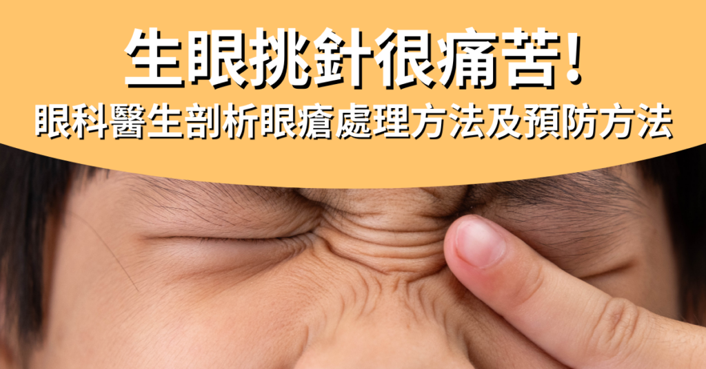
Peeping into someone else's bath will cause eye sores/eye needles? How long do symptoms take to get better? Is it possible to recover by applying ointment on your own? Or do you want to see an ophthalmologist for eye sores/eye needles? I believe that many people will have similar questions, especially the most want to know, once sick, how long will eye sores / eye needles be good? Before answering these questions, we need to understand the pathogenesis.
Causes of eye sores/eye needles
Eye sores, commonly known as eye needles, are medically called "styes". Eye sores/needles usually occur near the meibomian glands and can cause redness and swelling of the eyelids. The main cause of eye sores/needles is excessive oil secretion of the meibomian glands, resulting in blockage of the glands, coupled with the lack of attention to eye hygiene on weekdays, which causes bacterial infections.
In addition, if the patient often develops eye sores/needles, it may also be because the hair follicles of the eyelashes are blocked by mites. These tiny creatures can live at the roots of eyelashes, and when they grow in large quantities, they can lead to inflammation and the accumulation of pus, eventually forming eye sores/eye picks. So in fact, it is not formed because of peeking at other people's baths.
To prevent eye sores/needles, daily eye care and cleansing are very important, especially for people who wear makeup frequently, be sure to remove makeup thoroughly and avoid sharing makeup and makeup tools with other people as much as possible. If eye sores/needles are almost intolerant and tend to occur, an ophthalmologist should be consulted to ensure that the sores/needles condition is managed appropriately.
Characteristics and symptoms of eye sores/needles
Eye sores/needles grow in different locations and correspond to different names, namely "external styes" and "internal styes". The sebaceous glands that grow on the more superficial eyelashes are external styes; The internal styes grow in the deep meibomian gland, because the pus point is on the inside of the eyelid, so the inflammation time will be longer.
At the beginning of the disease, there is only a slight foreign body sensation, and it will not be painful to the touch, and such symptoms will heal themselves as long as they rest more and apply appropriate heat. However, if there is a bacterial infection with eye sores/needles, the following symptoms will occur:
眼挑針症狀
1. Symptoms of eye sores/eye needles
Slight redness and swelling of the eyelids
Minor itching and dandruff
There is a foreign body sensation
A scratching sensation when blinking
2. Severe symptoms of eye sores/eye needles
The eyelids are visibly red, swollen, enlarged, and even affect the field of vision
Pain, eyelids accompanied by slight warmth
The foreign body sensation is heavy
Transient astigmatism is present
If persistent swelling and pain are found, an ophthalmologist should be consulted immediately for eye sores/needles.
How are eye sores/needles treated?
Some patients try to pluck the affected area with a needle and squeeze the pus out when dealing with eye sores/needles, but this may cause bacterial infection, causing the wound to become inflamed or recur again, or causing cellulitis or even brain infection. Therefore, doctors do not encourage patients with eye sores/needles to be treated in this way. If you find that you have eye sores/needles, you should consult an ophthalmologist. Usually, when doctors treat and treat eye sores/needles, they will provide appropriate treatment recommendations according to the severity of the patient.
1. Mild eye sores/eye needles
To deal with mild eye sores/eye pick-and-needles, as long as you pay attention to eye hygiene, you can try to warm towels and "hot eggs" to warm the compress to make the eye sores/eye pick-needles subside on their own, the method is to boil the eggs, do not need to peel the shell, wrap them in a gauze towel, gently massage the eyes, apply 4 to 5 times a day, each time for 15 minutes, the blocked oil will melt and push out, but also reduce the blockage of the hair follicles by the oil, this method of eye sores / eye picks needles will be good without a few resistances.
However, if you find that the eye sores/needles are too painful, it is recommended to seek help from a doctor who will also prescribe some steroids or antibiotics according to the eye sores to help reduce inflammation and prevent infection.
2. Severe eye sores/eye needles
If you notice that the eye sores/needle are still inflamed and do not improve, the doctor will recommend that the person with eye sores/needle pick needle inject corticosteroids to remove the lump. However, this medication whitens the surrounding skin and is not optimal for dark-skinned people, so doctors may recommend minor surgery to remove eye sores/needles. This type of surgery is fairly simple and the wound is minimal, and there is no scarring on the appearance. If eye sores/needles still occur frequently in the same area after surgery, doctors will send the tissue to a laboratory to rule out cancer, but usually lumps on the eyelids are benign and harmless.
How good is eye sore/eye needle?
As for eye sores/eye needles, it depends on the severity of the patient. Generally speaking, mild eye sores/eye needles should be patient and wait a few days to get better, and the swelling will slowly subside; Severe patients need to be more patient, and eye sores will not improve until almost 2 weeks. If eye sores/needles are treated with resistance, they will get better in almost a few days and the course of the disease is expected to be shortened.
Whether it is a mild or severe patient, when dealing with eye sores/eye needles, the most important thing is to seek medical attention in time, and be patient to wait a few days to see if it will improve, self-treatment of eye sores/eye needles is not recommended, because this will cause more problems, resulting in the final eye sores/eye needles are almost more patient first will improve, recover, then may even take more than a whole month to recover.
Are eye sores/needles contagious?
In general, eye sores/needles are not contagious by themselves. However, if you do not treat eye sores/needles in time, which can lead to the growth of bacteria or hair follicle mites, there is a risk that it may be transmitted to the other eye or to others through contact. Sharing towels, pillows, or cosmetics with people increases the risk.
Will eye sores/eye needles be almost intolerant and often recur?
Anyone who has long sores or needles knows that the feeling is not only quite uncomfortable, but sometimes directly affects the appearance. Although this is not an emergency, it is definitely the biggest problem for some people, because eye sores/eye needles are almost intolerant and often recur. To prevent and prevent recurrence of crises, the most important thing is to start with hygiene, diet and preventive treatment.
Pay attention to hygiene: keep your hands clean, especially before touching your eyes; Also avoid touching your eyes with your hands and wiping your eyes with unclean towels
Healthy work and diet: reduce staying up late and let the eyes fully rest, in order to effectively regulate the oil secretion of the meibomian glands; Reduce the consumption of fried or spicy foods to reduce the chance of inflammation in the body
TREATMENT TO PREVENT RECURRENCE IS MORE EFFECTIVE IN PATIENTS WHO HAVE RELAPSES THAT ARE ALMOST INTOLERANT TO EYE SORES/NEEDLES, AS THEY KILL PARASITES, ARE ANTI-INFLAMMATORY, AND ANTIMICROBIAL. TAKING IPL (INTENSE PULSED LIGHT) AS AN EXAMPLE, IPL HAS PHOTOBIOREGULATORY FUNCTIONS, EFFECTIVELY STIMULATES CELL MITOCHONDRIA AND PROMOTES CELL HEALTH. TREATMENT WITH ANTIBIOTICS CONTAINING TETRACYCLINE OR MACROLIDES CAN HELP WITH ANTI-INFLAMMATORY AND BACTERIOSTATIC TREATMENT.
If not properly managed, eye sores/needles may be tolerant and often recur after a few months, or cause other more serious eye problems. Therefore, if there is eye sore/needle picking, it is best to seek an ophthalmologist to manage eye sores/eye needles.
Smoking: Studies have shown that smokers are 3 to 4 times more likely to develop macular degeneration than the general population
Poor eating habits: Long-term intake of high-fat, high-sugar and other foods causes obesity and three highs, affects the health of eye blood vessels, and increases the risk of macular degeneration
LONG-TERM EXPOSURE TO INTENSE SUNLIGHT: UVA IN THE ULTRAVIOLET RAYS OF SUNLIGHT ACCELERATES THE AGING OF PHOTORECEPTOR CELLS, CAUSING DAMAGE TO THE MACULA
CELL GROWTH FACTOR ROLE: SOME GENETIC ABNORMALITIES IN THE HUMAN BODY OR RELATED TO MACULAR DEGENERATION, SUCH AS COMPLEMENT FACTOR B, COMPLEMENT FACTOR H, THESE GENE ABNORMALITIES HAVE BEEN SHOWN TO BE GREATLY RELATED TO THE DEVELOPMENT OF MACULAR DEGENERATION
In addition, although blue light from electronic products is rumored to be one of the causes of age-related macular degeneration, there is not enough research data to confirm this claim. However, using electronics in a darker environment may cause the macula to absorb more blue light, which can lead to various eye diseases.
Smart low vision glasses project images of their surroundings in real time to the healthiest areas of the retina of low vision people and adjust them to the patient's perception. These glasses improve the patient's perception of real-world scenes by supplementing and correcting visual details. During the setup process, the ophthalmologist can intuitively adjust the glasses to help the patient find the best vision.
Bifocal additional intraocular lens
SML (Scharioth Macular Lens) is an implantable lens that can help patients "magnify" their field of view, with a specially designed central optical area that provides a high additional optical focal point of +10.0D to improve advanced vision without compromising distance vision.
Will some macular degeneration get better after surgery?
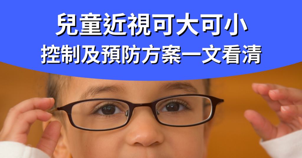
Myopia is one of the most common eye problems among children nowadays. According to the Department of Health, about 18% of children aged 6 in Hong Kong have myopia. The rate of myopia in 12-year-olds is as high as 62%, which is much higher than that in other Eurasia regions. In addition to bad eye habits, if parents themselves have high myopia, the chance of children suffering from myopia will also increase significantly. Therefore, early prevention and control of myopia in children is extremely important.
Electronic products are the leading cause of myopia in children
With the popularization of electronic products, the age group of myopic children has gradually increased. Many parents use electronic products as children's pacifiers, and children develop the habit of relying on electronic products when their eyes are not fully developed, affecting the normal development of their eyes.
Among children with myopia, up to 80% of children myopia is caused by poor eye habits. Watching the electronic screen for a long time will make the pupil constantly adapt to the change of light source, so that the ciliary muscle that regulates the pupil is always in a state of tension, resulting in excessive eye fatigue and enhancing the risk of myopia in children. To prevent or control myopia in children, parents must strictly control the amount of time their children spend using electronic devices, encourage them to participate in outdoor activities, and reduce their reliance on electronic nipples.
Is myopia in children also true and false?
But in fact, myopia also has "true myopia" and "pseudomyopia", the former is because the eye structure has changed, so it cannot recover naturally; The latter is a manifestation of temporary eye fatigue, which can be improved as long as the eye muscles are properly relaxed. Since the two types of myopia recover in different ways, the focus should be on understanding which type of myopia children belong to.
Pseudomyopia often occurs in children under the age of 12, because the ciliary muscles of children are quite powerful, and they use electronic products for a long time in close proximity, which is prone to the inability of the ciliary muscles to relax naturally, resulting in pseudomyopia. However, as long as the eyes are sufficiently rested, children with pseudomyopia can return to normal vision.
If you suspect that the child at home has vision problems, it is recommended to seek an ophthalmologist for examination immediately, usually pseudomyopia in children can be restored, most of them are ordered to relax the eyes, so as to detect the true refractive state of the eyes, to judge true/false myopia.
Although pseudomyopia in children can recover, it is also a major sign of myopia, and if children do not correct their eye habits in time, they may also become true myopia.
How to find out if a child has myopia?
Children will not realize that they have vision problems, so parents should pay extra attention to whether children have myopia or other vision problems through observation. If the following signs of myopia occur, parents should take their child to consult an ophthalmologist:
Blink or rub your eyes frequently
Often pull the corners of your eyes or squint to see things in the distance
Often tilt your head to see things
When looking at things, the eyes are close to things
What are the consequences of neglecting to control myopia in children?
Some parents may think that if their children's myopia is not deep, they can ignore it. However, in fact, if children's myopia is not controlled, the degree of myopia will gradually increase with age, and even soar in a short period of time. If you already have myopia at a young age, it is easy to cause myopia to exceed 500 degrees.
But do you think it's just as simple as deepening myopia? No! When the degree of myopia is deeper, the chance of suffering from eye diseases such as cataracts, glaucoma, and macular degeneration will increase exponentially in the future, and severe may even lead to blindness.
Cataracts: People with high myopia tend to degenerate and cloudify their lenses, leading to an increased risk of cataracts.
Glaucoma: People with high myopia have higher intraocular pressure and are 4 times more likely to develop glaucoma.
Macular degeneration: Among patients with myopia of 800 degrees and myopia for 10 years, about 5~10% of patients have macular degeneration.
Floaters: The vitreous of highly myopic people is prone to degeneration, resulting in fibrofilamentous suspended solids.
Retinal detachment: The retina of highly myopic people is prone to degeneration or atrophy, and the risk of developing retinal detachment is more than 20 times that of ordinary people.
Therefore, parents should actively pay attention to the degree of myopia in children and take control and preventive measures to avoid children becoming highly myopic in the future.
What are the ways to control myopia in children?
Generally speaking, the ideal rate of myopia progression for children should be maintained at no more than 50 degrees per year. If children's myopia increases by more than 100 degrees per year, it is an unusual situation, and it may develop into high myopia in the future, and even increase the risk of complications, so the medical community has been committed to developing a variety of methods to control and improve myopia in children in recent years to slow down the progression of myopia in children.
Low-concentration atropine eye drops
Low-concentration atropine eye drops can inhibit scleral overgrowth, slow down eye elongation, and then control the progression of myopia in children, which can slow the progression by 67% in 1 year.
However, some children may experience photophobia side effects after using low-concentration atropine eye drops, or may not be clear when seeing things at close range, so caution should be taken before using low-concentration atropine eye drops.
ORTHOKERATOLOGY CONTACT LENSES (OK LENSES)
ORTHOKERATOLOGY CONTACT LENSES (OK LENSES) ARE CONTACT LENSES WORN AT NIGHT AND ARE SUITABLE FOR CHILDREN WITH MYOPIA WHOSE MYOPIA DOES NOT EXCEED 600 DEGREES. THE PRINCIPLE IS TO HELP SHAPE THE CORNEA THROUGH HIGHLY OXYGEN-PERMEABLE RIGID LENSES, ALLOWING USERS TO SEE CLEARLY DURING THE DAY WITHOUT GLASSES. STUDIES HAVE CONFIRMED THAT LONG-TERM WEARING OF OK LENSES CAN CONTROL THE DEEPENING OF MYOPIA IN CHILDREN, AND GENERALLY CAN DELAY THE SPEED OF MYOPIA IN CHILDREN TO 50%~80%.
However, orthokeratology contact lenses are only temporary vision corrections, and if they are stopped, the deepening of myopia in children will return to their original state.
IN ADDITION, THE PRICE OF OK LENSES TO CONTROL CHILDREN'S MYOPIA IS USUALLY AROUND HK$5,000~HK$10,000, PLUS THE LENSES NEED TO BE REPLACED EVERY 9 MONTHS OR SO, SO THE PRICE OF SUCH CHILDREN'S MYOPIA CONTROL LENSES MAY BE A BIT BURDENSOME FOR SOME FAMILIES.
DEFOCUSING LENSES (DIMS)
There are two degrees of defocusing lenses, namely ordinary myopia lenses in the center and peripheral lenses, which use myopic defocusing optical elements to focus peripheral images in front of the retina, thereby slowing down eye elongation and controlling the speed of myopia in children. Studies have confirmed that children who wear defocusing lenses have significantly reduced myopia by 52% within 2 years.
UNFORTUNATELY, DEFOCUSING LENSES ARE ONLY SUITABLE FOR MYOPIC CHILDREN WITH MYOPIA OF NO MORE THAN 650 DEGREES AND ASTIGMATISM OF NO MORE THAN 400 DEGREES, AND CHILDREN WHO EXCEED THIS DEGREE CAN ONLY FIND OTHER CONTROL SOLUTIONS. IN ADDITION, THE PRICE OF THESE CHILDREN'S MYOPIA CONTROL LENSES IS NOT LOW, AND MOST OF THE COMMERCIALLY AVAILABLE CHILDREN'S MYOPIA CONTROL DEFOCUSING LENSES START FROM HK$4,000, WHICH DIRECTLY DISCOURAGES PARENTS WHO WANT TO USE LENSES TO CONTROL CHILDREN'S MYOPIA.
AN EARLIER METHOD TO CONTROL MYOPIA IN CHILDREN - AI MYOPIA CONTROL PROGRAM FOR CHILDREN
As mentioned above, some eye drops to control myopia in children may have side effects, and the current high price of defocusing lenses for controlling myopia in children, coupled with the so-called "prevention is better than cure", parents are beginning to realize that to control children's myopia and the chance of myopia deepening, they should not start planning after discovering that they have myopia, but should start to control the risk of myopia in children at an earlier stage.
THE CHILDREN'S AI MYOPIA CONTROL PROGRAM IS A KEY RESEARCH AND DEVELOPMENT PROJECT IN HONG KONG IN RECENT YEARS, WITH A PREDICTION ACCURACY RATE OF MORE THAN 80%. THE PURPOSE IS TO PREDICT THE DEVELOPMENT OF MYOPIA IN CHILDREN IN THE NEXT 3 YEARS IN ADVANCE, AND CUSTOMIZE INDIVIDUALIZED TREATMENT PLANS IN ADVANCE TO CONTROL THE DEEPENING OF MYOPIA OR RESTORE VISION IN CHILDREN.
THE MAIN PROCESS OF THE CHILDREN'S AI MYOPIA CONTROL PROGRAM IS AS FOLLOWS:
Fill in the questionnaire on children's habits
Perform various eye examinations
Enter the examination data and analyze the children's myopia in the next 3 years through artificial intelligence and big data
Parents and ophthalmologists can use the reports and charts to find out whether children's myopia development is at high risk
Based on the evaluation results, the ophthalmologist will customize personalized treatment for the child to delay the progression of myopia in children and reduce the risk of high myopia.
At present, the children's AI myopia control project led by ophthalmologist Dr. Tang Wenjie has been verified in 129,242 adolescent populations, with a prediction accuracy rate of more than 80%, providing early warning of myopia among adolescents.
PARENTS CAN ACCURATELY PREDICT THEIR CHILDREN'S FUTURE MYOPIA CHANGES THROUGH THE "AI INTELLIGENT PLATFORM FOR MYOPIA CONTROL IN CHILDREN", AND THE PREDICTION RESULTS CAN BE INTELLIGENTLY ANALYZED THROUGH THE BIG DATA OF THIS AI PLATFORM, SO THAT PARENTS CAN OBSERVE THE CHANGES IN THE REFRACTIVE ERROR PERFORMANCE OF THEIR CHILDREN AT DIFFERENT AGES ACCORDING TO THE PREDICTION REPORT (PARENTS CAN SEE THIS AS THE "REFRACTIVE DEVELOPMENT FILE"). BASED ON THE FORECAST REPORT, THE OPHTHALMOLOGIST WILL ALSO ANALYZE THE CURRENT MYOPIA SITUATION OF THE CHILDREN FOR PARENTS IN DETAIL, COMPARE THE NORMAL REFRACTIVE PERFORMANCE OF THE SAME AGE GROUP, AND FORMULATE PERSONALIZED MYOPIA TREATMENT PLANS.
Everyday ways to improve or control myopia in children
To prevent or control myopia, in addition to seeking the help of an ophthalmologist, parents can also help myopic children recover or improve their vision from daily life.
Establish proper eye habits
Try to stipulate that children should maintain a correct sitting posture when doing homework or reading, avoid lying on their stomachs or lying down, and should also avoid them reading or playing with mobile phones in a dim environment, and control the daily use of electronic products by myopic children, and develop the good habit of resting for 5~10 minutes every 30 minutes with their eyes.
Consume eye-friendly foods
PARENTS CAN ALSO GIVE THEIR CHILDREN MORE EYE PROTECTION FOODS SUCH AS VITAMINS A, C, E, LUTEIN, ANTHOCYANINS, ETC., WHICH CAN HELP MYOPIC CHILDREN RESTORE OR IMPROVE THEIR VISION.
Outdoor activities
It is recommended that parents take children to outdoor activities every day, such as playground play, park walking, reduce their time to use electronic products, and outdoor sunlight can also stimulate the secretion of retinal dopamine, effectively control the deepening of myopia in children.
Myopia in children develops very quickly, and the slightest attention can quickly soar. Therefore, in addition to supervising children to develop good eye habits in daily life, it is recommended that parents regularly take children for eye examinations. Once abnormalities are found, appropriate control measures and treatment plans can be taken in time to ensure the health of children's vision.
Presbyopia is a vision problem that occurs with age, and almost everyone has presbyopia, usually in people around the age of 40. Many people with presbyopia are reluctant to wear reading glasses for fear of being ridiculed for their age, which affects their daily lives. However, now that there are more and more ways to correct presbyopia, wearing reading glasses is not the only option, such as the Presbymax presbymax laser surgery highly recommended by ophthalmologist Dr. Tang Wenjie, which can not only avoid the embarrassment of wearing reading glasses, but also restore the vision of the eyes when they were young.
What is presbyopia?
The most common symptom of presbyopia is dyslexia, reading the text on the mobile phone or newspaper can become quite strenuous, and it is necessary to constantly adjust the distance to find a clearer focus. Prolonged close movement can make it easy to feel eye strain, dryness or discomfort; When working at close range in darker environments, more light is needed to help the eye focus.
The main cause of presbyopia is the degeneration of the lens and ciliary muscle in the eyeball. This degradation causes the lens to become stiff and inelastic, which in turn affects the eye's refractive ability and ultimately vision.
Although presbyopia usually occurs in people in their 40s, however, with the advancement of technology, people are increasingly relying on electronics. Staring at the screen for a long time will cause the ciliary muscle to tighten too much, causing the eyes to enter the presbyopia stage early. Therefore, protecting eye health is extremely important for modern people. It is recommended to reduce the time spent using electronic products to allow enough time for the eyes to relax to slow down the development of presbyopia.
2. Method self-test for presbyopia
Method 1: Symptom check
The following are some common symptoms related to presbyopia, if you get more than 3 of them, you may suffer from presbyopia.
When looking at the phone, you need to hold farther away to see clearly
When looking at the subject, I want to take off my glasses, but I can see more clearly without glasses
Feel that your home or office is darker than before
Often misread numbers or words
Eye strain is often felt
Shoulder and neck aches, stiffness
Often frowning, dizziness
Method 2: Banknote test
Take out a note and place it where you can see it best
Then focus on the small print on the banknote
Look away from the banknote and look at an object 2 to 3 meters away for about 5 seconds
Then look back at the small print and see if you can read the small print in a few seconds
Being able to focus within 1 second is normal; More than 1 second is at risk of presbyopia; More than 3 seconds can basically determine that there is presbyopia.
If you suspect that you have presbyopia, it is recommended to consult an ophthalmologist as soon as possible for professional advice and help. Your ophthalmologist will give you the need to wear reading glasses based on your vision needs or recommend Presbymax laser surgery to improve presbyopia. Early recognition and treatment of presbyopia can help improve vision and improve quality of life.
What is the difference between presbyopia and farsightedness?
Presbyopia and farsightedness are also blind, but the causes of the two are not the same. In addition, if you find that you have vision problems, it is recommended to go for a test.
Presbyopia is the degeneration of the eyeball
Presbyopia is an age-related vision problem in which the lens and ciliary muscle of the eye gradually deteriorate with age, making it less focused and making it difficult for the eye to see clearly in close objects and dark environments. If presbyopia has seriously affected your life, Presbymax presbyopia correction surgery can be considered. This is because Presbymax can not only treat presbyopia, but also help improve myopia, farsightedness, astigmatism and other problems, and restore the eyes to their youthful state.
Farsightedness is innate
Farsightedness is a vision problem related to the structure of the eyeball. It is mainly formed by short eyeballs or weakness of the ciliary muscle. If only the eyeball is short but the ciliary muscle is developed, there will basically be no problem of blurred vision; When the ciliary muscle is weak, it needs to be forced to focus on the near object. Farsightedness is innate, meaning that most children have farsightedness, but there is no need to worry too much, as the eye develops normally, the farsightedness will be relieved.
Leave the old flowers alone, be careful of physical problems!
Many patients feel that presbyopia has little impact on life and are reluctant to face up to the problem of presbyopia, thinking that they can see clearly by simply squinting, and even reading glasses are reluctant to wear. However, this behavior will cause ciliary muscle overwork, increase the burden on the eyes, cause eye soreness and discomfort, eye pain, etc., and in severe cases, it will even spread to the shoulder and neck area, resulting in shoulder and neck pain, headache, dizziness, nausea and other symptoms.
Although presbyopia is not an emergency, the degree of presbyopia will increase with age. If presbyopia at 100 degrees is diagnosed at 40 degrees, it will increase to 300 degrees at 60 degrees, and the degree of presbyopia will increase by about 10 degrees per year. If the presbyopia degree decreases abnormally, this may be a warning sign for cataracts, and patients should seek detailed examination and appropriate correction from an ophthalmologist as soon as possible. Timely identification and treatment of presbyopia is the key to protecting eye health.
What are the ways to correct presbyopia?
With the continuous advancement of medicine, more and more ways to correct presbyopia, including monofocal glasses, multifocal lenses, contact lenses or surgical implantation of intraocular lenses, Presbymax presbyopia laser surgery, etc.
Monofocal glasses: Usually monofocal glasses only need to be worn when looking at things up close. Such glasses can only provide clear vision at a specific distance. However, if a person has both myopia and presbyopia, then it is necessary to frequently remove and wear two glasses of different degrees in different situations, and then multifocal glasses can be considered.
Multifocal glasses: These glasses are equipped with multiple lenses with different focal points, allowing you to see far, medium and close distances in one pair of glasses without the need to change glasses frequently.
Multifocal contact lenses: If you don't like the bulkiness of glasses or people who are used to wearing contact lenses, you can use multifocal contact lenses or directly have surgery to correct your vision.
Surgical treatment: surgical treatment can include monocular vision surgery, multifocal intraocular lens surgery, Presbymax presbyopia laser correction surgery, etc. Presbymax presbyopia laser correction surgery is a relatively new option. This procedure uses laser technology to reshape the cornea to improve presbyopia vision, thereby reducing or eliminating dependence on glasses or contact lenses.
Traditional vision correction surgery VS Presbymax presbyopia correction, which one to choose?
Monocular vision surgery
Multifocal intraocular lens
Presbymax presbyopia
❌ Medium-range images are blurry
❌ Poor three-dimensionality
❌ There is a glare problem
❌ Not suitable for night motorists
❌ It takes time to get used to
✅ Far, medium and close vision is clear
✅ No glare issues
✅ No time is required to adapt, and normal activities can be carried out 24 hours after surgery
Presbymax presbyopia surgery mainly uses laser polishing on the surface of the cornea to make the cornea multifocal state. The main operation is to correct the myopia, farsightedness, astigmatism, etc. of the main eye to close to 0 degrees; The other eye retains or creates myopia of about 150 to 250 degrees, which is dedicated to seeing close distances and improving presbyopia. In addition, Presbymax also adds Spherical Aberration to increase the depth of field of both eyes, extend focus, and change chromatic aberration, thereby creating a 'fusion zone' for both eyes, allowing patients to obtain clear vision at the same time, and ensure the original color and stereoscopic acuity.
Dr. Tong said that Presbymax is one of the most recognized and mature presbyopia correction methods in the world. Patients who have undergone Presbymax presbyopia surgery are extremely satisfied and the price is relatively affordable. The procedure is very short, about 10 minutes to complete, within 24 hours the patient can move normally, within a few weeks vision can be fully restored, no discomfort, so Presbymax can be the best choice for presbyopia.
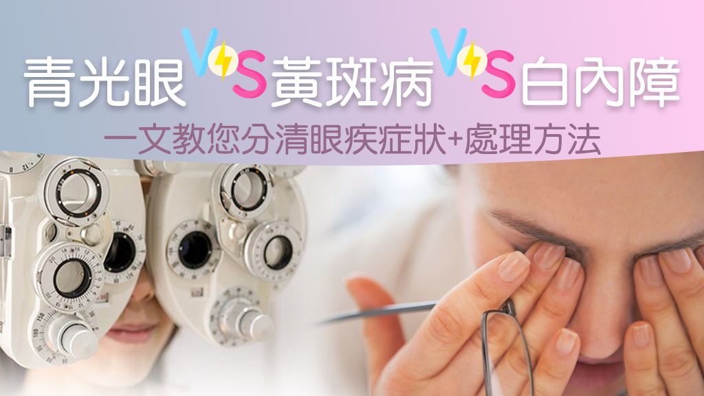
According to the World Health Organization, about 39 million people worldwide are blind, of which 51% are caused by cataracts, the leading cause of blindness in the world. The most common cataract is senile cataract, which is usually found to have cataract symptoms in people over 50 years old, while diabetics and deep myopia are more likely to develop cataract symptoms earlier. If the cataract becomes mature, it will bring a series of complications to the patient, such as: glaucoma, uveitis, etc., and even blindness, with serious consequences. Therefore, patients will undergo cataract surgery, treat cataracts with intraocular lenses, and restore clear vision to the eyes through treatment.
What is Cataract?
Cataract is caused by the transparent lens used to focus in the eye becomes cloudy due to old age or disease, and the lens functions like a camera lens, helping the eye to focus accurately, so that the image can be focused on the retina to form a clear image. The lens is made of proteins and has the function of regulating light. When the lens becomes cloudy, the transparency decreases, and light cannot pass through, resulting in decreased vision and cataracts.
What are the symptoms of cataracts?
The symptoms of cataract are relatively easy to identify compared with other eye diseases, such as dry eye, and the most noticeable symptoms generally include reduced reading ability, which is mainly caused by blurred vision, and there will be photophobia problems, patients are also prone to glare at night, or symptoms such as loss of color and contrast resolution. The symptoms of cataract can be subdivided into early cataract symptoms and late cataract symptoms:
Initial cataract symptoms
photophobia
Monocular diplopia
Myopia suddenly spiked geometrically
Presbyopia lightened
Late cataract symptoms
Vision modeling
See things glare
The image color becomes dull
Why do I get cataracts?
aging
The main cause of cataract is due to aging of the eyes due to aging, which is also known as "senile cataract", which is caused by the aging and decline of the lens. In medicine, according to the opacity location of the lens, such cataracts are subdivided into three categories: posterior subcapsular opacity, nuclear opacity, and cortical opacity.
Caused by other diseases
Also known as "secondary cataracts", it is caused by surgery, steroid medications, or other health problems such as glaucoma, diabetes, intraocular inflammation, and high myopia.
Caused by trauma
Trauma is also a cause of cataracts. When the eye has been subjected to strong impact, puncture injury, or has been subjected to high heat, chemical burns, etc., the crystal fibers are misaligned, and lesions such as turbidity and hardening may occur. However, some patients do not develop cataracts immediately, but gradually appear later.
Open surgeryto treat cataracts is the only way out
At present, open surgery is the only treatment for cataracts. The cataract surgery process involves removing the cloudy lens and implanting an intraocular lens to treat the cataract and improve vision. Generally, cataract surgery is performed under local anesthesia, and doctors use a microscope in the operating room to remove the originally cloudy lens and implant an intraocular lens suitable for cataract patients using ultrasound emulsification technology. Since this is a minimally invasive procedure, the surgical incision will be relatively thin and stitches will not necessarily be required, so the wound recovery time is relatively short. Cataract surgery is safe and complications are uncommon.
However, since everyone's condition and needs are different, and intraocular lenses are not as easy to replace as glasses, you should understand with your doctor before undergoing cataract surgery, and then choose the cataract intraocular lens that suits you.
Before undergoing cataract surgery, the patient decides which intraocular lens to choose for cataracts. Patients can decide which intraocular lens for cataract treatment can bring the best results for patients based on their overall eye health, such as dry eye, corneal astigmatism or macular health, etc., and finally undergo cataract surgery.
At present, there are three main types of intraocular lenses available for cataract surgery:
Unifocal intraocular lens
This is a commonly used intraocular lens in cataract patients and is also suitable for all patients. However, this intraocular lens only has a single focal length, and if the single-focal IOL lens implanted with the long-distance viewing function improves farsightedness, it may be necessary to wear reading glasses when performing close activities such as reading or playing sparrows.
All-round wide view of intraocular lenses
Lengthen the eye focus, solve the shortcomings of the previous multifocal length intraocular lens that can only see far and close things, and fill the mid-distance vision.
Trifocal intraocular lens
This intraocular lens is suitable for cataract and presbyopia patients, it is a multifunctional intraocular lens with far, medium and near full vision, and after surgery, cataract patients will have less dependence on glasses. However, cataract patients may have visual disturbances such as halos and glare after surgery, so patients may need more time to adapt.
In addition, the new generation of continuous intraocular lenses launched in recent years provides a wide continuous field of view from a distance to a reading distance of 33 cm, eliminating the visual gap existing in trifocal technology and other multifocal technologies, allowing patients to experience continuous high-contrast vision from far to near even under low-light conditions, greatly reducing the dependence of cataract patients on glasses. Its UV light filtering technology also helps to reduce glare and halo intensity when driving at night.
Many patients who undergo cataract surgery to treat cataracts will question the duration of the IOL. In fact, current IOLs are made of biocompatible materials such as acrylic or silicone. This means that they do not react with the human body anaphylaxis. For more than 100 years, no cases of aging intraocular lenses affecting vision have been found. So, don't worry about intraocular lens life. IOLs live longer than anyone can live, and rare IOLs change and need to be replaced.
When should I have cataract surgery?
Because when cataracts become mature and worsen, there is a chance to bring a series of complications to patients, such as glaucoma, uveitis, etc., so early treatment should be received cataract surgery. Moreover, under the current medical practice, patients do not need to wait for the symptoms of cataract complications before surgery. Cataract surgery can be considered when vision gradually deteriorates and vision is insufficient to cope with daily work or daily needs. After the patient's vision loss is detected by the nurse, after examination and discussion with the doctor, the decision can be made when to undergo surgery to treat cataracts.
If you wait until the advanced cataract for surgery, the lens will expand or even dissolve, which will bring a series of complications to the patient, such as glaucoma, uveitis, which can lead to irreversible blindness, and advanced cataract surgery is more difficult and vision recovery is slower.
Care aftercataract surgery
Patients may have glasses, sunglasses or eye protection for one week after cataract surgery. Do not wipe your eyes with your hands to avoid inflammation of the wound. The eye area can also be gently rubbed with disinfectant cotton or clean paper towels to avoid infection. Normal movement after routine cataract surgery without bed rest. However, complex cases such as trauma and concomitant glaucoma may require restriction of activities.
In terms of diet, abstinence is not required after surgery, but patients should reduce their intake of irritating foods. For people with allergies, foods with high protein content should also be avoided.
Do not swim, do strenuous exercise, and avoid lifting or carrying heavy objects over 30 pounds for one month after surgery. On weekdays, when the head is bent low, be careful to avoid collisions.
Have you ever tried to work for a while, but you feel that your eyes are dry and itchy, and you can't help blinking, even if you close your eyes and rest? This may be a symptom of dry eyes! Want to improve dry eyes, think that just eye drops, artificial tears, or reduce the TV and computer screens, can improve dry eye symptoms from the root cause of the disease? If you think it won't take long for dry eyes to heal on their own, you're very wrong! It may be that these methods will only make the symptoms of dry eye more and more serious.
What is dry eye?
Dry eye is a common eye disease, with 3 out of 10 Hong Kong people having dry eye. It occurs when the eye can't produce enough tears, or when the tears aren't working properly, which can make the eyes feel dry and itchy. The cause of dry eye syndrome is mainly because with age, tear secretion decreases, clinical data show that by the age of 65, the secretion of the lipid layer in tears is reduced by 60% compared with 18 years old, tears evaporate faster, so dry eye patients themselves are mostly older groups.
How does the composition of tears affect eye health?
Before we know how to improve dry eye symptoms, we should know how tears can do in eye protection. In addition to keeping the cornea moist and providing optical properties, tears can also provide oxygen to the cornea, and also have the effect of washing away impurities and foreign bodies entering the eye, while the antibodies and enzymes can resist foreign bacteria and protect the health of the cornea.
The tears on the surface of the eye are made up of three layers, each of which plays a specific role, and if there is a problem with one of the layers, the tear cannot moisturize the eye completely, causing dry eye syndrome.
First, the grease layer
The outermost layer is the oil layer, which is lubricated by the secretory glands under the eyelashes, which not only prevents the aqueous components from evaporating too quickly, but also increases the surface tension to maintain the stability of the tear layer while providing lubrication of the eyelids.
Second, the water layer
The aqueous layer is the intermediate layer, the main component of tears, produced by the lacrimal glands. It is an important component that provides nutrients to corneal epithelial cells and contains anti-infective substances that protect the eye from bacterial infection.
Third, the mucin layer
The innermost layer is the mucin layer, secreted by conjunctival cells, and its main function is to contact the cornea and conjunctival epithelium, providing lubrication and ensuring that the water layer adequately wets the cornea.
The composition and function of these three layers of tear fluid must be kept adequate, and it needs to be evenly distributed over the eyes by blinking to keep the eyes comfortable. If one of the layers is undersecreted or unevenly distributed, the eyes will experience a feeling of dryness, sometimes even foreign body eye pain, tingling or temporary hazyness, and may even cause dry eyes.
What are the subjective symptoms of dry eye?
Have you ever felt dry eyes, itchy, foreign body sensation, photophobia, and blurred vision? Or do you feel that the tears in your eyes are less secreted than ordinary people? However, do these symptoms mean dry eyes?
In fact, the symptoms of dry eyes are not a disease, but eye discomfort caused by external factors. As for xerophthalmia, most patients will have the following subjective symptoms, including:
Dry eyes
Foreign body sensation
Burning sensation
Pins and needles
Itchy eyes
photophobia
jealous
Fatigue easily
Blurred vision
Vision fluctuations
The reason for the above symptoms is that patients with dry eye have poor tear layer stability and are prone to many of the symptoms mentioned above. The reason is that the discomfort of dry eye stimulates the lacrimal glands, causing them to secrete a large amount of reflex tears, while making the patient very sensitive to wind and light, and often temporary blurred vision.
How to objectively tell if you have dry eye?
TO ACCURATELY DIAGNOSE WHETHER YOU HAVE DRY EYE, THE MAIN THING IS, OF COURSE, BASED ON A SERIES OF SYMPTOMS AND TEST RESULTS. HOWEVER, IF YOU ARE SIMPLY SUSPICIOUS AND WANT TO KNOW IF YOU HAVE DRY EYE, YOU MAY WISH TO SELF-TEST FOR DRY EYE THROUGH AN INTERNATIONALLY RECOGNIZED OCULAR SURFACE DISEASE INDEX (OSDI).
First, the eye sensation in the past week
anytime
Most of the time
About half the time
Occasionally
not
Dry
4
3
2
1
0
photophobia
4
3
2
1
0
Redness with bloodshots
4
3
2
1
0
ache
4
3
2
1
0
Foreign body sensation
4
3
2
1
0
Thick discharge
4
3
2
1
0
Blurred vision
4
3
2
1
0
Poor vision
4
3
2
1
0
2. The following activities have been affected by eye discomfort in the past week
anytime
Most of the time
About half the time
Occasionally
not
not applicable
Read
4
3
2
1
0
NA
Use a mobile phone/computer
4
3
2
1
0
NA
Night driving
4
3
2
1
0
NA
Watch TV
4
3
2
1
0
NA
3. In the past week, the eyes have not felt comfortable in the following conditions
anytime
Most of the time
About half the time
Occasionally
not
not applicable
When the wind blows (eyes are afraid of the wind)
4
3
2
1
0
NA
Dry environment
4
3
2
1
0
NA
Air-conditioned room
4
3
2
1
0
NA
Dry eye syndrome scoring method
OSDI = (sum of scores x 25) / sum of answers
OSDI SCORE
Symptoms.
0~12
No, normal
13~22
Mild dry eyes
23~32
Moderate dry eye
33~100
Severe dry eye
If you do this self-test and find that your score is about 13 points or higher, you may need to seek a more accurate test. To confirm the diagnosis of dry eye, ophthalmologists usually evaluate it with an ocular tear film analyzer (OSA) or a tear secretion test (Schirmer's test).
1. EYE SURFACE TEAR FILM ANALYZER (OSA)
THE SURFACE TEAR FILM ANALYZER (OSA) IS AN ADVANCED EXAMINATION METHOD THAT ACCURATELY DETECTS ALL ASPECTS OF THE EYE, INCLUDING THE STRUCTURAL FUNCTION OF THE TARSAL LINE, THE THICKNESS AND ANALYSIS OF THE TEAR FILM OIL LAYER, TEAR RUPTURE TIME, TEAR RIVER HEIGHT, AUTOMATIC BLINK RECORDING, AND QUANTIFICATION OF GLAND DISRUPTION.
Tear secretion test (Schirmer's test)
Schirmer's test is a common method to measure tear secretion, a special filter paper is placed on the lower eyelid for about 5 minutes, according to the wetness of the filter paper can know the basic secretion of tears, the normal value should be more than 10 mm. The tear film rupture time refers to the time when the tear film completely ruptures after the patient blinks, and the normal value should be more than 10 seconds.
According to the above criteria, if the patient has only eye symptoms, but there are no changes in tear secretion during eye examination, it is dry eye. If it is only a simple dry eye, it can be relieved after ordering eye drops, and it is not a dry eye syndrome, it may just be a condition of visual fatigue, such as temporary eye discomfort after reading books for a long time, reading the computer, dry eyes, photophobia and tears, blurred vision and other phenomena.
What causes dry eyes?
There are many causes of dry eye, mainly related to the state of the tear film and the function of the lacrimal glands, including insufficient secretion of tears, or poor quality of tears, so dry eye syndrome can be divided into two categories:
Dehydrated dry eye
Evaporative type of dry eye syndrome with oil deficiency
Many people mistakenly think that dry eyes are dehydration, avoiding dry eyes can improve dry eyes, but in fact, the lipid layer of the outer layer of the eye can prevent the evaporation of the tear film, and the lipid layer secretes clear oil by the meibomian glands, locking the tear film formed on the surface of the protective eyeball, so that the tear fluid is not easy to evaporate and keep the eyes moist. When the oil secreted by the patient is thick or even solidified, it is easy to cause tears to evaporate quickly, which in turn makes the eyes feel dry, which is also the cause of dry eyes. Therefore, the cause of dry eye syndrome is not necessarily water shortage, but also oil deficiency, that is, the cause of dry eye syndrome can be subdivided into four types: insufficient secretion of water layer, imperfect secretion of oil layer, uneven distribution of tear film, and improper secretion of mucin layer.
First, the secretion of the water layer is insufficient
Insufficient secretion of the water layer is the most common cause of dry eyes. This may be due to the reduced function of the tear glands due to aging, and women are also prone to dry eyes after menopause, which may be related to changes in hormone secretion in the body. In addition, some autoimmune diseases (rheumatoid arthritis, systemic lupus erythematosus), blood diseases (lymphoma, leukemia), trauma, infection, autonomic nervous disorders, long-term use of certain eye drops or drugs, etc. can also cause insufficient tear secretion. Long-term wearing of contact lenses can also affect the secretion of tears, because contact lenses reduce the sensitivity of the cornea, reduce tear secretion, and dry eyes will also occur.
Second, the secretion of oil layer is not perfect
Inadequate oil secretion is also a common cause of dry eyes. This is usually due to eyelid disease causing poor function of the eyelid sebaceous glands, which in turn affects the oil layer of the outer layer of the tear film, so that the tear cannot be effectively maintained on the surface of the eyeball, resulting in dry eye syndrome.
Third, the tear film is unevenly distributed
Excessive evaporation of tears and uneven distribution of tear film are also causes of dry eyes. Eyelid disease can lead to poor eyelid closure, excessive evaporation of tears. In addition, prolonged concentration on driving, staring at the TV, using the computer and other activities will reduce the number of blinks, which in turn will affect the distribution of tears. Prolonged work in an air-conditioned room or in a hot and dry environment may also affect the stability of the tear film, resulting in dry eyes.
Fourth, the mucin layer is secreted improperly
THE MUCIN LAYER IS THE INNERMOST LAYER OF THE TEAR FILM, WHICH IS SECRETED BY CONJUNCTIVAL CELLS AND HAS A LUBRICATING EFFECT TO MAINTAIN AN EVEN DISTRIBUTION OF TEARS ON THE SURFACE OF THE EYE. VITAMIN A DEFICIENCY, CHRONIC CONJUNCTIVITIS OR PEMPHIGOID DISEASES CAN LEAD TO INSUFFICIENT MUCIN SECRETION, WHICH IN TURN AFFECTS THE STABILITY OF TEARS. IN ADDITION, WHEN THE EYES ARE EXPOSED TO CHEMICALS OR HARMFUL SUBSTANCES, IT CAN LEAD TO DAMAGE TO CONJUNCTIVAL CELLS, WHICH IN TURN AFFECTS THE NORMAL SECRETORY FUNCTION OF THE MUCIN LAYER.
Who is at high risk of dry eye?
Dry eye syndrome has many causes, from age, living environment, autoimmune diseases and daily habits.
1. People over 50 years old
Aging is one of the main causes of dry eye, as aging lacrimal gland function and hormonal imbalance can lead to insufficient tear production, and between the ages of 50 and 55 is at high risk for dry eye.
Second, long-term wear of contact lenses
When using contact lenses for a long time, it may interfere with the normal secretion and distribution of tears, and it is easy to produce dry eye syndrome, especially soft contact lenses, wearing watery soft contact lenses such as eyes is like putting a sponge that is very absorbent in a place with water, it will suck away all the nearby water.
Third, long-term use of electronic products
Long-term use of electronic devices such as smartphones and tablets reduces the frequency of blinking, which is easy to cause insufficient tear secretion and prone to dry eyes.
4. Driving for a long time
Air conditioning and ventilation systems in vehicles dry out the air, which in turn accelerates the evaporation of tears, leading to dry and uncomfortable eyes. At the same time, staring ahead for a long time and paying attention to traffic conditions will also keep the eyes focused for a long time, reduce the number of blinks, and further increase the risk of dry eyes.
5. Unhealthy eating habits
Omega-3 fatty acids are important for maintaining healthy tear production and reducing eye inflammation. When a diet deficient in omega-3 fatty acids, such as a low intake of fish (salmon, mackerel, sardines), flaxseeds, chia seeds, and walnuts, increases the risk of dry eye.
6. Patients with chronic diseases or eye injuries
Studies have found that people with conditions such as diabetes, glaucoma, thyroid disease and high blood pressure are more likely than others to develop dry eye syndrome because these chronic diseases may affect the production or composition of tears, which can lead to dry and uncomfortable eyes. In addition, if the eye has been injured in the past, such as trauma or surgery, it may lead to decreased or damaged tear gland function, because the repair process after an eye injury affects the secretion of tears and the protective layer on the surface of the eye, thereby increasing the risk of dry eye.
What are some ways to improve dry eyes?
Many patients want to know how long it will take for dry eye symptoms to get better and what can be done to improve dry eye symptoms immediately. However, to improve dry eye, it depends on the symptoms and the extent of the condition of the individual patient. At present, most of the medical profession will adopt artificial tears, autologous serum, thermal pulse therapy, pulsed light therapy and other methods to help patients improve dry eye syndrome.
1. Artificial tears
Patients with mild symptoms can improve dry eye syndrome by supplementing traditional artificial tears as a treatment method, including liquid type, gel type and ointment type, so as to improve dry eye syndrome. The use of artificial tears can reduce dry eye symptoms, avoid corneal damage, and maintain the smooth surface of the eyeball. However, sometimes the effect of artificial tears may not improve immediately and significantly, and most people with dry eye syndrome take longer to improve. However, depending on the individual, some patients may only take a few days, but more dry eye patients will take longer to improve, which may take weeks or even longer.
Second, autologous serum
In recent years, autologous serum has been found to contain components closer to tear fluid, so it has begun to be used in different ocular surface diseases, including the improvement of more severe dry eye patients. When artificial tears do not relieve symptoms, your ophthalmologist may recommend using autologous serum eye drops. However, the production of autologous serum eye drops is complicated, which requires the patient's blood to be drawn, then centrifuged, isolated serum, and finally made into eye drops of appropriate concentration and frozen in aliquots. These serums contain growth factors that have anti-inflammatory and healing effects. A single blood draw can usually make 6 to 10 eye drops, and because they do not contain preservatives, care must be taken to avoid contamination of eye drops, and it will take longer than most treatments, but it can also improve the symptoms of dry eye syndrome and allow patients to live a comfortable life again.
3. Thermal pulsation therapy
Some eye conditions are more severe and can be treated with Lipidflow. By using the instrument, warm at 40°C for 12 minutes, and massage the meibomian glands with regular pulsation, soften and unblock the blocked meibomian glands, improve dry eye symptoms, and avoid further atrophy caused by meibomian gland blockage, thereby improving eye health.
4. Pulsed light therapy
Intense Pulsed Light (IPL) is suitable for all levels of dry eye and can open up blocked tarsal plates, help normalize oil production, and reduce tear evaporation. The number of treatments usually takes 3 to 4 times, and the dry eye will gradually improve.
Are dry eye care methods effective?
To improve eye fatigue and dry eye syndrome, many people will choose supplemental health foods, such as lutein. However, although lutein can help filter blue light and prevent retinopathy, it does not help much to improve dry eye syndrome.
In addition, many people think that dry eyes can be cured with eye drops. However, eye drops can only temporarily relieve symptoms, and excessive use of eye drops can hinder the natural production of tears in the eyes, leading to dry eyes.
How can dry eyes be prevented or improved?
Instead of trying the above methods casually, it is better to protect your eye health and stay away from dry eyes through correct eye habits and avoiding bad habits.
First, the correct eye habits
When using electronic devices regularly, working or reading, you should take regular breaks from your eyes. One of the widely recommended methods is the 20/20/20 Eye Protection Rule, which rests your eyes for 20 seconds every 20 minutes while shifting your gaze to a distance of about 20 feet (about 6 meters) away from you. This helps reduce the stress associated with prolonged close use of the eyes and helps keep the eyes healthy.
Second, maintain a good environment
When working or resting indoors, special attention should be paid to the humidity in the air to avoid the adverse effects of too dry an environment on the eyes. In addition, we should also avoid direct exposure to the direct blowing airflow of the fan or air conditioner and adjust the angle of the fan or air conditioner to avoid blowing directly into the eye area or, where possible, away from direct contact with the airflow.
3. Supplement antioxidant nutrients
Antioxidant nutrients are essential nutrients that protect the eyes, which need light to become imaging. Consuming foods rich in omega-3 fatty acids, such as salmon, walnuts, and flaxseed, can reduce inflammation and stabilize the tear film. At the same time, consuming foods rich in vitamins A and C, such as carrots, oranges and kiwifruits, can help fight free radical damage and protect eye tissue.
4. Supplement anti-inflammatory phytochemicals
Dry eye is often associated with inflammation in the eye, and anti-inflammatory substances can help reduce inflammation in the eye, reduce discomfort, and promote the repair of eye tissue, thereby improving dry eye syndrome. Many phytochemicals have anti-inflammatory effects, such as quercetin, turmeric, anthocyanins, eugenol, etc., which can help improve dry eyes.
Floaters are sometimes annoying, people with floaters will see "black spots" floating in their eyes, not painful or itchy and unable to rub open, when you look directly at them, they will disappear in an instant. Floaters come in different shapes, such as black dots, lines, circles, ovals, tadpoles, etc., especially when looking at a bright and clean background, it is easier to spot its presence. Floaters are a degenerative disease of the eyes, mild patients in general, as long as they are used to coexisting with black spots and floating objects in the eyes, daily life is not affected, they do not need to receive floaters treatment, but if the black spots or floating objects in the eyes are too large or too much, floaters suddenly attack or flash, so that vision is damaged, you should seek improvement methods, find an ophthalmologist for the treatment of floaters for consultation and surgery or laser treatment of floaters, otherwise, the longer it drags on, the more serious or blind!
What is floaters?
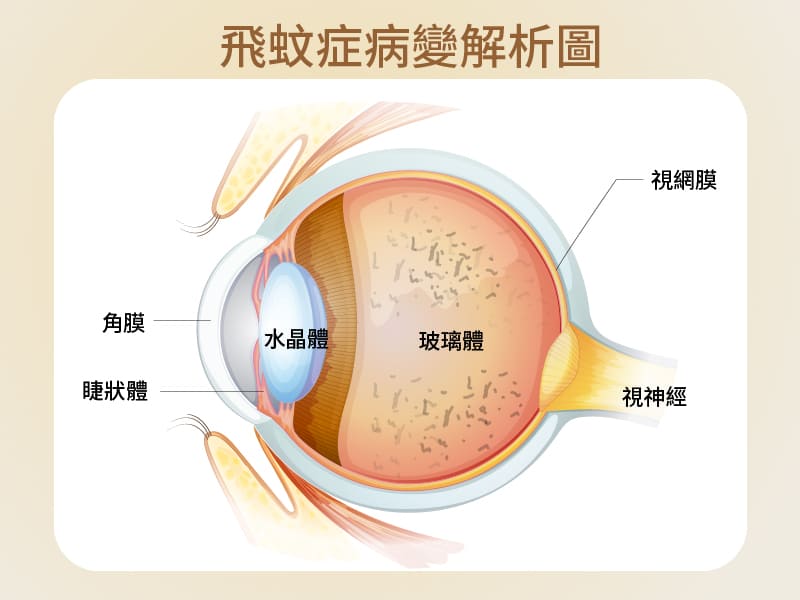
Floaters are medically a phenomenon of vitreous degeneration of the eye. The vitreous is a transparent gel located behind the lens and before the retina. Normally, the vitreous fills the entire vitreous cavity to support the shape of the eyeball. But with age, myopia and other problems, the vitreous will liquefy and shrink, forming turbid fibers, and these fibers will float around in the vitreous, when the light refracts these fibers, the patient will feel that there are black spots in the eyes, which is the precursor of floaters.
Floaters are medically a phenomenon of vitreous degeneration of the eye. The vitreous is a transparent gel located behind the lens and before the retina. Normally, the vitreous fills the entire vitreous cavity to support the shape of the eyeball. But with age, myopia and other problems, the vitreous will liquefy and shrink, forming turbid fibers, and these fibers will float around in the vitreous, when the light refracts these fibers, the patient will feel that there are black spots in the eyes, which is the precursor of floaters.
Who is most likely to develop floaters?
Floaters usually become more common with age, so older people are more likely to have dark spots in their eyes. But in addition to age, other factors that may increase the risk of floaters include myopia, history of eye surgery, eye trauma or eye inflammation. According to clinical studies, high-risk groups for floaters include:
Middle-aged and elderly
People with high myopia
People with hypertension/diabetes
Have had eye surgery
Blows to the head, e.g. car accidents, divers
Other eye problems, such as eye inflammation
However, most floaters are benign, such as because of old vitreous degeneration, or excessive eye fatigue, there may be symptoms of floaters, as long as there are more black spots in the eyes, and the position is fixed, do not worry too much.
Conversely, if suddenly there are a large number of dark spots in the eye that have affected the field of vision or there is a flash, it may be vitreous peeling, resulting in pulling the retina to the hole or even falling off. This condition must be investigated immediately, for improvement, or for floaters.
Causes of floaters
The causes of floaters can be broadly divided into three types, namely physiological, degenerative and pathological.
Physiological
About 80% of floaters are physiological. Physiological floaters usually occur in people under the age of 40 or people who have been using their eyes for a long time, and most people will see black spots in the eyes, which are vitreous impurities that usually do not affect vision, and do not need to be treated immediately for floaters, and will disappear on their own over time
Degenerative
As we age, the vitreous body of the eyeball, like other organs in the body, gradually deteriorates. In this degeneration process, the vitreous body will shrink to form fine fibers, these fibers will float in the glass, when the light enters the eye, and these impurities refract, it is the black spots in our eyes.
Pathological
Pathological floaters are caused by eye disease or systemic vascular disease. The so-called eye disease refers to the retina under the pull or degeneration of the process of the retina in the process of tearing or degeneration, resulting in vitreous hemorrhage, and even retinal peeling, resulting in visual impairment, and in the worst case, permanent blindness. In addition, systemic vascular diseases, such as eye stroke, diabetes, hypertension or macular degeneration, if not paid attention to and actively treated, may also appear pathological floaters, patients should seek to improve floaters as soon as possible.
Complications of floaters – retinal detachment/tear
Although suffering from floaters is not a serious disease, retinal detachment caused by floaters is an emergency in ophthalmology. One in four people with floaters may have their vision affected by retinal detachment or a tear. Keep in mind that if you have these signs of retinal detachment due to floaters, including:
There are a large number of dark spots in the eyes in a short time
Unusual flashes
shadow
The view is blocked
This means that there may be cracks in the retina, and it is important to seek immediate medical attention for treatment, otherwise floaters will cause permanent damage to vision or even blindness.
How is floaters treated?
If you have benign floaters, you do not need to be treated immediately. When you notice dark spots in your eyes, you can try moving the eyes to let the fluid flow through the eyes so that the fibers leave the line of sight. Of course, some people can't bear the black spots floating around in their eyes, and their attention is often disturbed by black spots, which seriously affects their mood, so they can consider floaters treatment.
With current technology, the main treatment and improvement of floaters is laser therapy and vitrectomy.
First, laser treatment of floaters
This floater treatment is suitable for large and concentrated vitreous fibers, using laser light to break these fibers into smaller fragments, through these methods to improve and eliminate floater symptoms. The laser treatment is performed under local anesthesia without pain and the whole treatment takes only 15 to 20 minutes. However, not everyone is suitable for laser treatment to improve floaters, and if the fibers are loose or too close to the macula or crystals, it should not be performed to avoid complications such as cataracts or macular damage.
Second, vitrectomy surgery to treat floaters
Vitrectomy is performed by removing the vitreous inside the eye through a small incision and replacing it with a solution to maintain the shape of the eye. The procedure only takes about 10 to 15 minutes. However, vitrectomy does not necessarily completely remove the fibers, and if the surgery itself causes bleeding or retinal tears, new floats may form, so most physicians do not recommend this procedure to treat floaters.
Floaters improvement and maintenance methods
Once floaters occur, it is almost difficult to reverse, even after laser or surgical treatment, there is a possibility of recurrence, what we can do is to live peacefully with it, or we can improve floaters by the following methods to reduce symptoms slightly.
First, the daily maintenance of benign floaters
Avoid excessive eye use
PAY ATTENTION NOT TO OVERWORK THE EYES, ESPECIALLY 3C HEAVY USERS, YOU SHOULD LET THE EYES REST FOR 5 TO 10 MINUTES EVERY 1 HOUR, USE THE REST TIME TO BLINK, LOOK INTO THE DISTANCE TO RELAX THE EYES.
Good living habits
In daily life, it is recommended to reduce the time spent playing with mobile phones, especially to avoid staying up late at night to look at mobile phones, to ensure that you have enough sleep and do exercise in a timely manner, which is also a way to improve floaters.
Regular follow-up
Do not think that you have benign floaters can relax, floaters must be regularly checked to avoid other lesions, miss the best time for treatment.
Eye care diet
Foods with antioxidant effects, such as berries, green or yellow vegetables, carrots, soybeans, milk, and fish oil, should be consumed in your diet to help improve floaters.
2. Postoperative care and maintenance of malignant floaters
Medication treatment
After surgery, be sure to follow the doctor's instructions, remember to wash your hands thoroughly with soap before each medicine to avoid cross-infection.
Improve your lifestyle
Rapid eye movements and excessive eye use should be avoided for 1 week after surgery. Try not to read books, mobile phones, computers, TV, etc. during this period. In addition, it is recommended not to do strenuous exercise, climbing, diving, or flying, as these activities can irritate the eyes and affect the speed of postoperative repair.
Use an eye patch
During eye repair, it is recommended to wear an eye mask at all times to prevent collisions and avoid unconsciously rubbing your eyes when sleeping at night. It is necessary to ensure that the eye mask is clean when using, and it is recommended to wash and disinfect the eye mask daily to avoid wound infection due to bacteria.
Daily diet
In addition to avoiding irritating foods, it is also necessary to avoid eating legumes, because such foods are easy to let the nitric oxide in the blood run into the eyes, causing the inert gas in the eyes to produce flatulence and increase the pressure in the eyes.
Safe at home
Keep your home clean and tidy, reduce dust and avoid secondhand smoke, and do not change the layout of the home or place objects in the aisle during the recovery period, so as not to cause collisions or falls in unfamiliar environments and irritate the eyes.
Regular follow-up visits
In addition to following the doctor's advice for regular follow-up consultations after surgery, if you notice eye discomfort, such as persistent eye pain, sudden blurred vision, nausea, increased or larger dark shadows, etc., remember to consult your ophthalmologist immediately to avoid worsening of the condition.
How can floaters be prevented?
Although we cannot completely avoid the occurrence of floaters, we can slow down the aging of the eyes and reduce the chance of developing floaters through daily maintenance.
1. Develop good eye habits
AVOID EXCESSIVE USE OF 3C PRODUCTS (1 HOUR WITH EYES AND 5-10 MINUTES BREAK)
Works in sufficient light and stable without flickering
Avoid staying up late to increase the burden on your eyes
2. Intake of eye care nutrients
Carotenoids (dark green, dark yellow and red fruits and vegetables)
Anthocyanins (plum fruits)
VITAMINS (VITAMINS A, B, C AND E)
Omega3 fatty acids (fish oil)
3. Proper control of systemic vascular diseases (such as: diabetes, hypertension)
Maintain a balanced nutrition and reduce the intake of foods high in oil and salt and sugar
Regular exercise (jogging, cycling)
Have regular full-body examinations
Although most floaters can not be treated, they should not be completely ignored, and they must be checked regularly, and if symptoms are aggravated, they should seek medical attention immediately to avoid missing the golden period of floater treatment.
Elderly people often have eye blindness, and may think that it is a degenerative problem that makes it difficult to see clearly, but have they ever thought that they may suffer from eye diseases due to eye lesions?
Eye disease symptoms
First of all, to distinguish whether vision suddenly or slowly decreased, visual impact is the middle range, the whole picture, or peripheral vision? And is there a feeling of pain in the eyes? So how to tell the difference between macular disease, cataracts and glaucoma? Taking cataracts as an example, the vision of the average elderly will slowly decline, and it will affect the whole picture. Macular disease, on the other hand, has a sudden and acute decrease in vision, affecting central vision. Glaucoma also slowly affects vision, generally affecting peripheral vision first.
These three diseases can be easily confused, and there are many ways for the elderly to check them. There is a kind of "checkered paper". Checkered paper is a kind of checkered paper, and the elderly can see whether the straight line has changed the curve by looking at the checkered paper to see if the straight line has changed the curve, and these conditions can determine whether there is macular disease. For other problems, an eye disease can only be determined through an ophthalmologist's examination.
How to heal?
There are generally two complications of the upper eye of diabetes, the main cause is hypoxia, resulting in angiogenesis. If angiogenesis occurs, those fragile blood vessels may have a condition that leaks, leading to vitreous hemorrhage or macular edema. To prevent this, it is usually necessary to inject eye drugs to prevent angiogenesis, thereby reducing hypoxia.
Cataracts are basically performed through minimally invasive cataract surgery, in which cloudy lenses are removed and implanted with intraocular lenses. Finally, glaucoma, with early treatment, the nerves can be controlled so that they do not continue to be damaged. There is no treatment for glaucoma, only testing, medication, surgery or laser to control intraocular pressure to slow down the damage to the nerves.
ELDERLY PEOPLE CAN PREVENT IT BY CHANGING THEIR DAILY LIFESTYLE, SUCH AS: LESS SUGAR AND LESS SALT; EAT FOODS RICH IN VITAMIN A, CAROTENE OR OMEGA FATTY ACIDS, BECAUSE OF ANTIOXIDANT EFFECTS; DO AT LEAST 30 MINUTES OF AEROBIC EXERCISE; DO NOT SMOKE. THE ELDERLY NEED REGULAR EYE EXAMINATIONS FOR EARLY PREVENTION AND EARLY TREATMENT. TRY TO AVOID ULTRAVIOLET RAYS TO IRRADIATE THE EYES TO PREVENT VARIOUS EYE DISEASES.