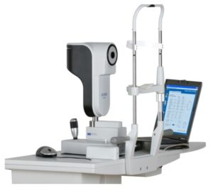
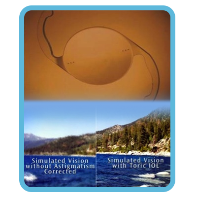
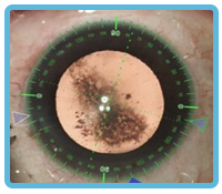
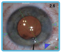
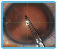
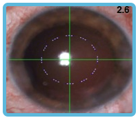
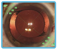
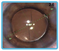
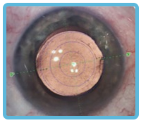
Surgery is the only way to treat cataracts. The surgical procedure involves removing the cloudy lens and implanting an intraocular lens to improve vision. Generally, cataract surgery is performed under local anesthesia, and the doctor uses a microscope in the operating room to remove the originally cloudy lens and implant a suitable intraocular lens using phacoemulsification technology. Since this is a minimally invasive procedure, the surgical incision will be thin and stitches may not be required, so the wound recovery time is relatively short. Cataract surgery is very safe and complications are uncommon.
Patients do not have to wait for cataracts to mature or for complications to develop before surgery. When the patient finds that the vision gradually deepens and the vision is not enough to meet the needs of daily work or life, the nurse will find that the vision has decreased, and finally decide when to undergo surgery after examination and discussion by the doctor.
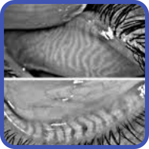
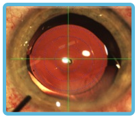
If the cataract becomes mature, when the cataract reaches an advanced stage, the lens will expand or even dissolve, which will bring a series of complications to the patient, such as glaucoma, uveitis, which can lead to irreversible blindness, and advanced cataract surgery is more difficult and vision recovery is slower.
The fee depends on the length and complexity of cataract surgery, including tools, instruments, drugs and consumables required for the operation. For details, please contact WhatsApp/by phone.