生眼瘡解決方法:熱敷藥膏都無效?割眼瘡手術幫你徹底解決 | 眼科醫生專業指南

How to treat stye: Are heat compresses and ointments ineffective? Eye stye surgery can help you get rid of it completely | Ophthalmology Guide

The eye infection has been recurring for more than two weeks; I’ve tried applying heat packs and applying ointment, but the swelling persists. Every day I look in the mirror and see my eyelids swollen and distorted, affecting my professional image and making it difficult for clients to see me without covering up.I have seen many patients in their 30s to 50s in my clinic who came seeking treatment after trying various home remedies and self-medication online; but in fact, they had delayed the best treatment time. When the eye infection persists and forms a hard lump, it indicates that it has entered the chronic stage, at which pointOnly an eye ulcer surgery can completely solve the problem.This outpatient minor surgery only takes 15 minutes, requires no hospitalization, and leaves minimal scarring, allowing you to recover quickly.

Why haven't your eye infections ever gotten better?

Many patients in their 30s and 50s come to the clinic and ask me the same question: "Why did my eye infections get better in a few days when I was younger, but now they persist and don't go away?" Actually, there is a very clear medical reason behind this.

As we age, our metabolism gradually slows down, the sebum secreted by the meibomian glands becomes thicker and more likely to clog up. Coupled with long hours of computer work, frequent late nights, and insufficient sleep in modern workplaces, these all reduce the immune system's function and weaken the body's ability to handle inflammation. So even though eye infections may resolve on their own in younger people, they become particularly persistent in this age group.

More importantly, many people mistakenly believe that eye infections are small problems caused by heat or lack of cleanliness, so they tend to self-treat for a while. But when you notice that the eye infection feels hard and not very painful but persists without resolving, it actually represents a transition from acute inflammation to chronic cyst (chloasma). In this case, the tissue within the cyst has become fibrous and hardened, and it cannot disappear on its own; it must be removed through surgery. This is also the real reason why so many methods you have tried have been ineffective.

Why are the viral eye infection remedies online not effective?

After discovering the eye infection, most people will first search online for solutions or ask friends for alternative remedies. Let me analyze the actual effectiveness of these common methods from the perspective of an ophthalmologist.

Heat compress method (hot chicken eggs, hot towel)

This is the most common method used online; there are even detailed instructions on how to apply it 4-5 times a day for 15 minutes each time. Theoretically, applying heat can help dissolve the blocked oil, but only if the eye infection is in its early stages and is only mildly swollen. The problem is that for busy working professionals, it is simply impossible to maintain this frequency every day. You have to rush to work in the morning, work at the office during the day, and then go home in the evening. It’s simply not possible. Moreover, if the eye infection has already formed pus or a chronic cyst, no matter how much heat you apply, it won’t dissolve on its own because the hardened tissue won’t dissolve on its own due to heat.

Antibiotic eye drops or ointments

Some people go to the pharmacy to buy eye drops or ointment, or after seeing a doctor, take the prescribed medication. Antibiotics can indeed control bacterial infections and prevent the situation from worsening, but they are only “controlling” rather than “healing.” For already formed abscesses or cysts, the medication simply does not reach the deeper lesions. I have seen many patients use the medication for 2-3 weeks, and the eye sore still remains swollen there; it is a waste of time and money.

Self-pressuring or tearing apart

This is the most unrecommended and dangerous approach. I know that when seeing pus-filled eye blisters, many people will involuntarily try to squeeze them out, hoping to solve the problem quickly. However, the blood vessels around the eyes are very complex and connected to the blood vessels in the brain. If squeezed by yourself, the bacteria may be pushed deeper into the tissue, and in severe cases, it can even lead to vasculitis or an infection in the brain.

Why aren't these methods effective for you?

If you have tried the above methods for more than 2-3 weeks without improvement, it is very likely that the eye infection has entered a chronic stage or is caused by a chalazion that is growing deep within the eyelid gland. At this point, continuing with conservative methods will only delay the condition; not only will it waste time, but you will also have to endure the discomfort and appearance problems caused by the eye infection every day. In this case,The eyelid surgery is the fastest, most effective, and most thorough solution.

Under what circumstances should eyelid surgery be considered?

When a patient presents with the following symptoms, I recommend that they seriously consider surgical treatment:

First, conservative treatment that lasts more than 2-3 weeks is ineffective.If you have tried heat therapy, medications, and other methods but your eye infection still hasn't subsided, this means conservative treatment is no longer effective for your condition; continuing to wait will only prolong the pain.

Second, the eye blisters are hard to the touch, not very painful, but they persist.This is a typical symptom of chronic cyst (sclerodermal cyst). The hardened cyst will not disappear on its own; it must be surgically removed. Many patients think that the painlessness indicates a recovery, but in fact, it is exactly the opposite; it indicates that the condition has entered a chronic phase.

Third, the eye infection continues to swell, beginning to affect vision or appearance.For working professionals who often have to deal with clients, meetings, and photo shoots, eye problems not only affect health but also seriously impact professional image and self-confidence. Instead of worrying about this every day, it is better to resolve the problem early through surgery.

Fourth, the same location keeps recurring.If your eye infection always occurs in the same area and you experience painful swelling every time, it means that the eyelid gland in that area is already damaged. Instead of allowing it to recur repeatedly, it is better to surgically completely clear the lesion.

What is the process of eye surgery?

Many patients are worried when they hear the word "surgery," but in fact, eyelid surgery is a very simple and safe outpatient procedure that may be completely different from what you imagine.

Basic information about the surgery:

  • Time: 10-15 minutes
  • Anesthesia: Local anesthesia
  • Hospitalization: Not required; you can go home the same day.
  • Wound size: 3-5 mm (the size of a grain of rice)
  • Scar: Almost invisible

The entire process is as follows: First, the medical staff will clean and disinfect your eyes. Then the doctor will inject local anesthesia into the eyelid area. At this point, you will feel a brief sting, but it will only last a few seconds. After that, you will not feel any pain during the entire surgical procedure; at most, there will be only a slight sensation of pressure.

After the anesthesia takes effect, the doctor will use specialized equipment to gently fix the eyelid, then make a small incision at the optimal location. If the chalazion is located on the outside of the eyelid, the incision will follow the skin texture and heal almost imperceptibly; if the chalazion is located on the inside of the eyelid, the incision will be made on the inside of the eyelid and will be completely invisible to the eye.

Next, the doctor carefully removes the pus, the contents of the cyst, or the inflamed tissue completely, ensuring that the wound does not bleed. Finally, an antibiotic ointment is applied, and the entire procedure is complete.

The anesthesia effect lasts after the surgery, so you won’t feel any pain. You can go home immediately and rest. There may be slight discomfort after the anesthesia wears off that night, but you can relieve it with ordinary painkillers.

How long after the surgery can I resume normal activities?

This is the most pressing issue for working professionals. Simply put:

Time phase Recovery status Notes
On the day of the surgery Half a day of time needs to be reserved. Including pre-operative preparation and post-operative rest
Day 2 You can work normally. Avoid overworking. If your work requires frequent meetings or client visits, your eyes may still be slightly swollen; you can wear glasses to cover them up.
Days 3-7 Normal work and life Take your medication on time, be careful to keep your wound clean, and avoid touching water. You can continue with your normal work and activities during this period, just don't engage in strenuous exercise.
7th-14th day Completely restored The follow-up visit allows the doctor to check on the recovery. Basically, she has already fully recovered and can wear makeup (but it is recommended to replace new eye makeup).

The entire recovery process takes about 1-2 weeks. The most important part is taking the medication as directed by the doctor, keeping the wound clean, and avoiding rubbing the eyes. After the surgery, it is important to eat a light diet, avoid staying up late, and give the body enough time to heal.

Why should I see an ophthalmologist?

Although eye infections are common, doctors can accurately determine the type of infection, assess whether surgery is really necessary, and choose the most suitable incision location and method. Follow-up after surgery is equally important. Doctors provide detailed post-operative care guidance, regularly schedule follow-up visits to assess recovery, and provide preventive recommendations based on your individual situation. This comprehensive treatment and follow-up can truly help you completely resolve your eye infection problem.

💬 Book an appointment now

Persistent eye problems? Book an appointment for evaluation and treatment options.


WhatsApp inquires about eye problems

Common questions FAQ

▶ 1. Will the eye surgery hurt a lot?

The surgery uses local anesthesia; the process is completely painless; there is only a brief tingling sensation during the injection of anesthesia lasting a few seconds. Mild discomfort after the procedure can be relieved with over-the-counter painkillers.

▶ 2. Will there be any scarring after the surgery?

Modern ophthalmic surgical techniques are sophisticated, with wounds measuring only 3-5 millimeters in size. They are also performed either along the skin texture or from the inside, leaving virtually no scarring after the procedure.

▶ 3. What is the approximate cost of the surgery?

Costs vary depending on the clinic and the individual case. It is recommended to inquire directly with the eye clinic. Some medical insurance plans may cover this surgical cost.

▶ 4. Will the infection return after the surgery?

Surgery can completely remove the lesion in that area, and the chance of recurrence in the same area is very low. However, if eye hygiene is not taken seriously, new eye infections may still develop in other areas, so post-operative care is important.

▶ 5. Do eye infections always need surgery?

Mild eye infections can be treated with heat therapy and medication initially. However, if conservative treatment is ineffective after 2-3 weeks or if a chronic cyst forms or recurrent episodes occur, surgery is the most effective solution.

▶ 6. Can people with diabetes have surgery?

Yes, but special attention should be paid to blood sugar control. Before the procedure, you should inform the doctor so that he or she can assess the risk and adjust the medication regimen.

▶ 7. How long after the surgery can I wear makeup?

It is recommended that you do not wear makeup for at least 1 week after the procedure, and that you replace all your eye makeup with new products to prevent bacteria from the old makeup from causing a new infection.


In conclusion: Don't delay anymore; timely treatment is the best solution.

While eye problems may seem like minor issues, if not treated properly or delayed, they can not only affect appearance and self-confidence, but also lead to more serious complications. For working adults aged 30 to 50, time is money; rather than spending every day worrying about eye problems and trying various ineffective methods, it is better to seek the help of an ophthalmologist as soon as possible.

The eyelid surgery is a simple, safe, quick, and effective treatment method. This small 15-minute procedure can completely solve the problem that has been troubling you for a long time, allowing you to regain clear vision and a professional appearance.

If your eye infection has lasted for more than 2 weeks, and you have tried both heat therapy and medication without success, or the recurring episodes are affecting your daily life, do not hesitate to contact us to schedule a detailed examination. We will provide a comprehensive assessment and recommend the most suitable treatment plan for you. Addressing the issue early can lead to a faster recovery.

How to treat stye: Are heat compresses and ointments ineffective? Eye stye surgery can help you get rid of it completely | Ophthalmology Guide Read More »

Understanding the risks and precautions of laser vision correction for presbyopia

1. 引言

隨著年齡的增長,人體會出現各種生理變化,其中視力的變化尤為顯著。老花眼是一種常見的自然生理現象,通常在四十多歲開始出現,顯著影響人們的日常生活。為了改善視力問題,越來越多的人選擇老花激光矯視手術。本文將深入探討這種手術的相關技術、費用、風險及注意事項,幫助大家更好地了解老花激光矯視。

2. 甚麼是老花激光矯視?

老花眼是由於眼晶體的彈性減少,使得近距離視力逐漸下降。老花激光矯視是利用激光技術調整角膜形狀,促使眼睛在不同距離的物體上獲得清晰的視覺。這種手術有助於減少或消除對老花眼鏡的依賴。

3. 老花激光矯視的治療技術

老花激光矯視的主要技術包括:

3.1 PresbyMAX老花矯視技術

PresbyMAX技術是一種針對老花眼的激光治療方法,與傳統的LASIK手術原理相似。此技術透過創建角膜瓣並進行激光處理,以形成多焦點的角膜形狀,幫助患者在不同距離的物體上都能獲得清晰的視力。

3.2 微笑矯視(SMILE)

SMILE技術是一種相對較新的激光矯視手術,它透過比傳統LASIK更小的切口來進行操作。這種技術具更小的手術傷口和較短的恢復時間,同時減少對眼睛神經的損傷,從而降低術後眼乾等後遺症的風險。

4. 老花激光矯視的後遺症與風險

4.1 常見的激光矯視後遺症

儘管老花激光矯視手術的成功率很高,但仍然存在一些風險和後遺症,包括:

  • 術後短暫視力模糊
  • Dry eyes
  • 短暫光暈或眩光現象

4.2 如何減少老花矯視的風險

減少手術風險的幾個指導原則包括:

  • 在手術前與醫生詳細討論病史及職業需求
  • 術後定期追蹤檢查,及早發現問題

5. 建議事項

醫生建議患者在選擇激光矯視手術前,應評估自身的健康狀況及視力問題,明白手術的風險和收益。如果有高眼壓或其他眼部疾病,則需謹慎考慮是否適合進行該手術。

6. 結論與未來展望

老花激光矯視手術雖然可以顯著改善視力及生活質量,但患者在決定是否進行該手術時,必須充分了解所有相關風險及後遺症,並選擇適合的技術與醫療機構。隨著技術的進步,未來可望出現更加安全、有效的治療方案,幫助更多人擺脫老花眼的困擾。

FAQ

  1. 老花激光矯視的手術效果能持續多久?

大多數患者在手術後可以享有長期的視力改善,但隨著年齡的增長,可能仍需佩戴老花眼鏡進行輔助。

2.激光矯視手術風險大嗎?

激光矯視手術的風險相對較低,而風險主要來自於個人眼睛的特點及手術後的護理。充分的術前檢查及選擇經驗豐富的醫生有助於降低風險。

Understanding the risks and precautions of laser vision correction for presbyopia Read More »

Precautions for cataract surgery for the elderly

1. 什麼是白內障手術

白內障手術是一種常見的眼科手術,主要用於移除眼睛內的混濁晶狀體,並用人工晶體取而代之。這項手術普遍適用於隨著年齡增長,視力逐漸減退的長者。透過手術,患者通常能夠顯著改善視力,恢復正常的日常生活。

2. 白內障手術的原因和成因

白內障的成因可以多種多樣,主要有年齡因素、糖尿病、高血壓及長期使用類固醇藥物等。

3. 白內障手術的流程

白內障手術通常在局部麻醉下進行,過程大約需要20分鐘,包括微小切口、超聲波乳化及植入人工晶體。手術當天即可回家,無需住院。

4. 長者白內障手術的特殊考量

對於年長患者,醫生會考慮到他們的整體健康狀況,並可能需要進行額外的檢查,以確保手術安全。此類患者還需要遵循特定的術後護理,如減少活動量、遵從藥物治療計劃等。

5. 白內障手術康復時間

大多數患者在手術後幾天內會感覺到視力的逐漸恢復,但完全康復通常需要數周時間。患者應避免夜間駕駛,遵循醫生的復診計劃,定期檢查眼睛。

6. 白內障手術費用與政府資助

白內障手術的費用因地區及醫療機構而異。在香港,患者可以透過公立醫院或診所獲得經濟實惠的手術選擇。

正在考慮白內障手術?用 3 分鐘做個人工晶體初步配對測試,了解最適合日常生活需要嘅晶體方向。

👉 Take the test now

FAQ

1. 白內障手術後的視力會變得更糟嗎?

一般來說,手術後視力會改善,但有少數患者可能出現後囊膜混濁的情況,這可以通過激光治療輕鬆解決。

2. 白內障手術後需要住院嗎?

不需要,通常患者在手術當天即可回家,無需留院觀察。

3. 患有其他疾病的患者適合手術嗎?

患有糖尿病或高血壓的患者通常也可以安全地進行白內障手術,但需要醫生根據病歷進行充分評估。

💬 Book an appointment for cataract evaluation

由眼科醫生為你進行全面評估,了解最適合嘅治療方案

WhatsApp inquiry

Precautions for cataract surgery for the elderly Read More »

What is conjunctivitis? An ophthalmologist explains in detail

Iritis is an eye disease that mainly occurs in the iris part of the eye. The iris is a colored tissue located around the pupil that is primarily responsible for controlling the pupil size and regulating the light entering the eye. When the iris becomes inflamed, it can cause symptoms such as eye redness, pain, and blurred vision. This condition may be associated with other eye or systemic diseases such as rheumatoid arthritis, autoimmune diseases, or infections. Early diagnosis and treatment are crucial for preventing visual impairment caused by iritis.

What is conjunctivitis?

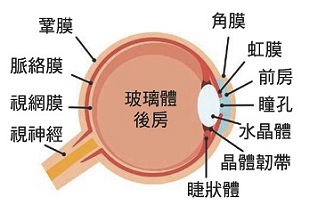
Retinal inflammation, also known as anterior uveitis or iridocyclitis, is characterized by the presence of noticeable red blood spots near the eye. It is usually caused by diseases such as infection, trauma, or immune system abnormalities. The iris is the part located between the cornea and the crystalline lens, which contracts or relaxes in response to the intensity of light to adjust the pupil size and control the intensity of light entering the eye. The iris has many blood vessels and pigments that nourish the eyes. When the iris becomes inflamed, the eyes become red and painful, and vision may also be affected. Retinal inflammation can be divided into acute and chronic types, and may also recur. Once symptoms of inflammation appear, it is essential to seek medical attention immediately to control the condition early on and reduce the severe complications that retinal inflammation can cause.

Are conjunctivitis and pink eye the same eye diseases?

For some people, conjunctivitis andRed-eye syndromeThe symptoms are also red eyes, so it is often mistaken for the same eye condition; only the name is different. In fact, although both conjunctivitis and red eye are inflammation of the eyes, the location of the inflammation and the symptoms are different.

As the name suggests, conjunctivitis occurs in the conjunctiva of the eye. If you observe closely, the blood vessels of conjunctivitis extend from the black eye towards the white of the eye. Red eye syndrome is also known as conjunctivitis, and when the conjunctiva becomes inflamed, the inner capillaries become swollen and swollen, causingThe eyes turn redUnlike that, in red-eye syndrome, the swelling usually spreads from the white part of the eye towards the black part.

Iris inflammationRed-eye syndrome (conjunctivitis)
SymptomsRed eyes, eye pain, glare, blurred vision, dilated pupils, increased floatersThe eyes are swollen, painful, and light-sensitive. There is tearing, foreign body sensation, itching, and the secretion is sticky white or yellow. In severe cases, there may be subconjunctival hemorrhage and damage to the cornea.
Treatment methodsSteroid eye drops, vasoconstrictors, immunosuppressantsAntibiotic eye ointment eye drops cold compress
Contagious無有

Types of conjunctivitis

Acute conjunctivitis

Acute conjunctivitis usually develops suddenly within a few hours or days, with obvious symptoms including red eyes, pain, and sensitivity to light. It usually affects only one eye. Acute conjunctivitis is usually not contagious unless caused by an infection. Doctors usually examine the anterior chamber and the cornea of the eye to diagnose the condition, and may perform imaging tests if necessary. Acute conjunctivitis requires immediate treatment to prevent rapid worsening of the condition. This type of conjunctivitis can also recur, especially in patients with autoimmune diseases.

Chronic conjunctivitis

Chronic conjunctivitis symptoms are generally mild at first, but they persist for a long time, and the disease usually progresses over a period of more than 3 months. Therefore, regular eye examinations are essential. Treatment for chronic conjunctivitis includes long-term use of low-dose steroid eye drops, and sometimes oral immunosuppressive agents are required. Like acute conjunctivitis, chronic conjunctivitis is usually not contagious. Both acute and chronic conjunctivitis can recur, so it is important to follow your doctor’s instructions regarding medication and regular check-ups.

Iris inflammation symptoms

  • Red eyes
  • Eye pain
  • Fear of light
  • Blurred vision
  • Pupillary distortion
  • Flying flies increased

Causes of conjunctivitis

Currently, the cause of most cases of conjunctivitis is unknown; doctors generally do not focus on what may be the underlying cause of the condition. However, if the conjunctivitis continues to recur, doctors may recommend blood tests or more detailed physical examinations to determine whether it is caused by an autoimmune disorder. The most common causes of conjunctivitis are as follows:

  • Autoimmune diseases:Conditions such as rheumatoid arthritis and ankylosing spondylitis may cause a disruption in the body's immune system, leading to conjunctivitis.
  • Infection:Infections with bacteria, viruses, or fungi can cause conjunctivitis, especially in patients with weakened immune systems.
  • Trauma:After an injury or surgery to the eye, inflammation of the iris may occur.

Complications after iridocyclitis

If left untreated, conjunctivitis can lead to conjunctival scarring that impairs the normal function of the pupil, resulting in blurred vision. Other more serious complications of conjunctivitis include cataracts, glaucoma, and corneal inflammation, and may even lead to blindness. Therefore, if you notice any signs of conjunctivitis in your eyes, you should seek immediate medical attention from an ophthalmologist for examination and treatment.

Treatment methods for iridocyclitis

Common treatments for conjunctivitis include eye drops, dilating agents, and immunosuppressants; however, the specific treatment method will be determined based on the patient’s condition. Ophthalmologists may combine two or more medications to help patients more effectively improve their conjunctivitis; therefore, please do not use over-the-counter prescriptions recommended online without medical advice. If you have any eye problems, please consult a doctor immediately.Consult with an eye specialist.That's right.

Steroid eye drops

The most common treatment method is the use of steroid eye drops to relieve conjunctivitis symptoms. Steroids can effectively reduce the inflammation of the conjunctiva, but they should be used under the guidance of a doctor to avoid side effects.

Dilating lens

Eye dilators can help enlarge the pupil and prevent the iris from sticking to the cornea. They also help relieve pain and photophobia, but long-term use may require monitoring of eye pressure.

Immunosuppressive agents

For chronic conjunctivitis caused by autoimmune diseases, immunosuppressive drugs may be needed to control the condition. These medications can reduce the immune system's attack on the conjunctiva and reduce the risk of inflammation.

Irritant food and drink prohibitions for conjunctivitis

It is recommended that patients should try their best.Foods that can boost the body's immune system should be avoided.This leads to an overactive immune system, which in turn attacks healthy body tissues or worsens the condition. Additionally, the patient also needs toReduce alcohol intake、High sugar、SpicyEat foods that stimulate the immune system to prevent exacerbating the inflammation of conjunctivitis. In addition to the doctor's prescription, you can also consume omega-3 fish oil, flaxseed oil, and foods rich in vitamins C and E in moderation on a daily basis to suppress and reduce eye inflammation.

Common problems

How long does conjunctivitis usually take to get better?

The recovery time for conjunctivitis depends on the severity of the condition and the treatment method. Acute conjunctivitis can usually be controlled within a few weeks, but chronic conjunctivitis may take several months or even longer to recover.

Can conjunctivitis be transmitted?

Conjunctivitis itself is usually not contagious unless caused by an infectious disease. In this case, contact with the source of infection may increase the risk of transmission.

Will the conjunctivitis recur?

Choroidal vasculitis can recur, especially in patients with autoimmune diseases. Therefore, regular eye examinations and continuous immunosuppressive therapy are crucial for preventing recurrence.

Should I refrain from drinking alcohol with conjunctivitis?

For patients with conjunctivitis, fasting and appropriate dietary adjustments help reduce inflammation. Avoiding high-sugar, high-fat, and spicy foods and consuming more foods rich in antioxidants helps maintain eye health.

Can conjunctivitis be cured?

Whether conjunctivitis can be completely cured depends on the cause that triggered the inflammation. If it is caused by an infection, once the infection is controlled, the inflammation may not recur. However, for conjunctivitis caused by autoimmune diseases, long-term treatment may be required to control the condition and prevent recurrence.


Can conjunctivitis heal on its own?

Some mild cases of conjunctivitis may resolve on their own, but in most cases medical intervention is required to prevent vision damage and other complications. Therefore, it is recommended that any symptoms of conjunctivitis be treated promptly to ensure proper treatment.

What is conjunctivitis? An ophthalmologist explains in detail Read More »

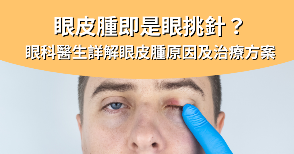
Is eyelid swelling the same as eye twitching? Ophthalmologist Explains the Causes and Treatment Options for Eyelid Swelling

Ephelides are a common condition; swelling can occur on both the upper and lower eyelids. Some are painless, while others are accompanied by red, itchy eyelids.

Common causes of eyelid swelling

There are many different causes of eyelid edema. The most common is edema. Possible causes of edema include drinking too much water before bed or consuming too much salt, which can lead to swelling. In addition, lack of sleep and stress can also be the cause of edema in the upper and lower eyelids, but these types of eyelid swelling usually do not cause pain or itching; they can usually be eliminated by making appropriate lifestyle and dietary changes.

However, in some cases, eyelid swelling may be caused by more serious factors. If you notice persistent swelling of the eyelids accompanied by pain, it is advisable to consult a doctor to determine the cause of the eye swelling and undergo appropriate treatment.

The following are some reasons why eye swelling may require medical intervention:

Eye allergies

Swollen eyes or a feeling of swelling may be caused by allergies to dust, pollen, certain foods, or medications. Eye allergies that cause swelling may also occur only on one side of the eyelid; this may be caused by contact with an allergen and then touching the single eye.

Cornoid cyst

Cornoid cystAlso known asEye pick needleThe main reason for this is blockage of the oil glands at the edge of the eyelids, which causes inflammation or bacterial infection, resulting in painful swelling of the inner and outer eyelids. The eyelids may appear noticeably swollen and the appearance may make the eyelids appear swollen.

Conjunctivitis

ConjunctivitisAlso known as red eye syndrome, it is usually caused by viruses, bacteria, or allergies. Conjunctivitis causes red eyes, burning and dryness, increased secretion, and painful sensation in the eyes and eyelids. Generally, patients with conjunctivitis are accompanied by swelling of the upper and lower eyelids, so it is recommended to seek medical advice from an ophthalmologist for appropriate treatment.

Blepharitis

Blepharitis can occur on the upper or lower eyelids. This inflammation causes the eyelids to become red, itchy, and painful. The causes of blepharitis include an overgrowth of Staphylococcus aureus, chronic skin inflammation, or prolonged failure to remove makeup, which can lead to allergic reactions in the eyelids.

Hyperthyroidism

Hyperthyroidism (HT) is a condition characterized by an autoimmune disorder. When the body’s immune system mistakenly identifies a healthy part of the body as a foreign invader, it releases antibodies to attack the healthy cells. HT attacks the outer surface of the eye, the eye cavity, and the eyelid tissue, leading to swollen eyelids, swollen and bulging eyes, and in severe cases, the risk of blindness.

Cellulitis

Sinusitis caused by a cold if not treated properly may lead to serious complications such as conjunctivitis. Patients with conjunctivitis may experience swollen eyes, red eyelids, burning and pain, and sudden visual impairment.

Insect bites

If the eyelid swelling is unilateral and accompanied by itching, blinking may cause some pain in the eyelid. This may be due to a mosquito bite. If you take good care of your eyes, the swelling will usually subside within a few days. However, there is also a possibility of further inflammation and swelling of the eyelid. It is advisable to monitor the condition at all times and seek medical attention if necessary.

Ways to get rid of eye swelling

Ice pack

Appropriate ice packs can help relieve the feeling of swelling and burning in the upper and lower eyelids caused by mosquito bites or allergies, but this does not have any therapeutic effect and can only temporarily alleviate discomfort.

Heat compress

Applying heat can promote blood circulation and also help soften and drain blocked oil or pus. Proper heat therapy can resolve eye problems such as hordeolum and eyelid inflammation.

Massage the eye area

Lightly pressing your index or middle finger around your eyes can help reduce swelling and relieve the itching sensation. It is important to note that you should thoroughly clean your hands before performing the massage to avoid eye infections caused by bacteria.

Drug treatment

Antibiotics, eye ointments, and eye drops can effectively control bacterial infections and the symptoms of inflammation and swelling of the eyelids. Since there are many causes of eyelid swelling, different causes and severity of symptoms may require different treatment options, such as antihistamines, thyroid medications, immunosuppressants, etc. Therefore, patients must follow their doctor’s prescription and instructions to properly treat the problem of eyelid swelling.

Surgical treatment

In cases of more severe conditions such as barley corn cyst, follicular inflammation, and hyperthyroidism, if the desired results are still not achieved after a series of treatments, the doctor will recommend that the patient consider surgical treatment to address the underlying problem of eyelid swelling.

In short, there are many causes of eyelid swelling; it is crucial not to take it lightly. If you notice persistent swelling and pain in your eyelids, it is recommended to seek immediate medical attention from an ophthalmologist for further examination and active treatment.

Common problems

How to get rid of eye bags?

The simplest way to relieve eyelid swelling is to apply ice or heat. In addition, maintaining good sleep and rest is also crucial. If you are unsure of the cause, you should still consult an ophthalmologist for an examination.

What should I do if my lower eyelid swells?

Under-eye puffiness may be caused by allergies, eyelid inflammation, or edema. It is recommended to use ice packs to reduce swelling and seek the help of an ophthalmologist to determine the cause.

Do I need to see a doctor for eyelid swelling?

If the eyelid swelling does not subside or is accompanied by other symptoms such as pain or visual impairment, it is recommended to seek medical attention immediately.

Why do the upper and lower eyelids suddenly swell but don't hurt?

Sudden swelling of the upper or lower eyelids that is not painful may be caused by minor allergies or infection; it may also be just water retention. If the symptoms persist or worsen, it is recommended to see a doctor for examination.

What to do if your eyelids are swollen when you wake up in the morning?

If your eyelids are swollen in the morning, it may just be simple swelling; it usually resolves on its own, and you can also apply ice to help reduce the swelling.

Is eyelid swelling the same as eye twitching? Ophthalmologist Explains the Causes and Treatment Options for Eyelid Swelling Read More »

眼角膜受損

Damaged cornea can lead to blindness! What should I do first?

The cornea is located at the front of the eyeball. Because it is exposed to the external environment, the cornea is more susceptible to damage. If the injured cornea is not treated carefully, the wound may not heal for a long time, leading to ulcers and even permanent visual impairment.

When we encounter wounds on the skin, we can use tape or ointment to promote wound healing; but if the cornea is unfortunately damaged, what should we do to avoid scarring or other complications?

What is the cornea?

The cornea is a transparent thin membrane located in the front chamber of the eye, in front of the iris and pupil, which is the frontmost part of the eye. Its main function is to allow light to enter the eyeball, thereby forming a clear visual image. The cornea not only prevents bacteria from entering the eye, but also has a highly developed sense of touch; if any foreign object touches the cornea, the eyelids immediately close to protect the eyes.

Causes of corneal damage

There are many causes of corneal damage; common ones include:

  • Foreign object intrusion:In case of eye injury caused by sand, a nail puncture, or other sharp objects, it may result in a corneal abrasion. After the cornea is injured, vision may immediately become blurred and the patient may experience pain. Additionally, accidental exposure to chemical substances can also damage the cornea, even leading to severe corneal ulcers.
  • Eye diseasesSuch as eyelash falling out, eyelid closure incomplete,Dry eye syndromeThis will all lead to painful eyes, stinging eyes, and damaged corneas.
  • Improper use of contact lenses:Using improper techniques to insert contact lenses into the eyes can easily cause damage to the cornea. Therefore, when wearing contact lenses, you should strictly follow the instructions and be careful when placing the lenses in the eyes. Ultraviolet rays from snow can also cause eye pain.
  • Misuse of eye drops:Long-term use of eye drops containing steroids may lead to thinning of the cornea and even perforation.
  • Ultraviolet radiation:Prolonged exposure to ultraviolet light (snow, sea surface) can cause death of the corneal cells and severe pain.

Symptoms of corneal damage

Symptoms of corneal damage include:

  • Vision decline:After the cornea is injured, vision becomes blurred; when looking at things, one may feel a transparent membrane covering the eye or a mist in the eye; the clarity of vision noticeably decreases.
  • Eye pain:Patients may experience painful or tingling eyes, especially when exposed to light.
  • Feeling foreign object in the eye:When the cornea is damaged, the eye may feel like there is a foreign object in it and have a blurry appearance, as if there were a transparent membrane in the eye.
  • Other symptoms may also include involuntary movements.Crying、ConjunctivitisWait.

Treatment for corneal injury

Mild corneal damage generally heals naturally within 1 to 2 days. However, when corneal damage occurs, prompt treatment is essential. If you experience severe discomfort in your eyes, it is recommended to immediately visit an ophthalmologist to treat the injury. Here are some common treatment methods:

  • Immediately rinse your eyes:If you experience a foreign body sensation or discomfort in your eyes, you should immediately rinse your eyes with eye drops to remove the foreign body from your eyes and avoid rubbing your eyes to prevent further damage to the cornea.
  • Drug treatmentDamage to the cornea is usually treated with eye drops; using antibiotic eye drops or artificial tears can reduce the risk of infection and help the cornea heal. If the patient continues to experience pain or the condition worsens, they should seek immediate medical attention.
  • Take proper rest:Reducing eye time spent using devices, especially avoiding excessive use of electronic devices, helps the cornea to self-repair.

Complications after corneal damage

If the eye is not treated promptly and appropriately after injury, serious complications may occur. Common complications of corneal damage include:

  • Permanent loss of vision:If scarring occurs, it may lead to a decrease in vision and may not be fully restored even after surgery.
  • Corneal ulcer:Untreated corneal injuries can lead to corneal ulcers, a potentially dangerous condition that can cause blindness.
  • Light-sensitive:After corneal injury, the eyes may become more sensitive, leading to an increased sensitivity to light.

Consult an ophthalmologist immediately.

Eye injuries can be minor or serious. If you experience any discomfort in your eyes, you should consult and have an examination with an ophthalmologist as soon as possible to allow a professional ophthalmologist to develop an effective treatment plan to maintain eye health.

Common problems

How long does it take for the cornea to heal after an injury?

How long it takes for the corneal abrasion to heal depends on the extent of the damage. Generally, minor corneal abrasions may heal within a few days, while more severe corneal injuries may take several weeks to fully recover.

Can corneal damage heal on its own?

Yes, corneal damage has some self-healing ability. Mild damage usually heals within a few days, but severe damage may require medical intervention. Using appropriate eye drops for corneal damage can also promote healing.

Will corneal damage recover?

In most cases, corneal damage can be completely restored; especially mild damage. However, if severe sequelae or corneal ulcers occur, it may lead to permanent visual impairment.

How to know if the cornea is damaged?

If you experience a foreign body sensation in your eyes, blurred vision, or stinging pain, this may be a sign of damage to the cornea. If you experience sudden decrease in vision or symptoms such as sensitivity to light, the possibility of corneal damage should also be considered.

Will the cornea heal within a few days after being injured?

The healing time for corneal injuries is usually between a few days and a week. Mild injuries usually heal quickly, but severe injuries may take longer and should be treated by a professional doctor.

Will the cornea regenerate?

Yes, the cornea has some regenerative ability. The corneal epithelial cells usually regenerate quickly after injury, thereby repairing minor abrasions. However, deeper corneal damage may require a longer recovery time and may leave scarring that affects vision.

Damaged cornea can lead to blindness! What should I do first? Read More »

弱視_封面圖

There is a golden period for treating low vision; early detection can improve the healing rate.

弱視,又稱為「懶惰眼」,是兒童常見的視覺發育問題之一。它主要發生在視覺發育關鍵期內,由於各種原因導致眼睛無法正常地傳遞視覺信息到大腦,從而影響視力的正常發展。弱視如果不及時治療,可能會導致永久性視力損傷,因此及早發現並進行治療至關重要。

弱視定義

弱視通常在嬰兒/兒童時期出現,是指一隻或兩隻眼睛基於眼睛發育不良、大腦缺乏視覺刺激導致視力低於同齡兒童正常水平。

如果弱視兒童在戴眼鏡或隱形眼鏡矯正後依然無法達到同齡應有的標準,或兒童兩眼視差二行以上(如0.5和0.7),都會被確診為懶惰眼。

弱視症狀

通常來講,弱視從肉眼是無法看出異樣的,但我們可以從孩子的日常表現中察覺。幼兒弱視(懶惰眼)的特徵有:

  • 看不清事物,如閱讀或看電視時需要靠得很近
  • 孩子經常出現鬥雞眼/習慣性閉上一隻眼睛看事物
  • 孩子經常歪頭/瞇眼
  • 眼睛深度感、立體感異常,難以判斷物體的距離,導致經常跌倒
  • 眼睛對光敏感
  • 雙眼協調不良,表現為斜視,孩子的一隻眼睛可能會偏左/右或上/下

弱視成因

屈光性弱視

當兒童有眼睛屈光不正(High myopia、遠視或散光)的問題,又沒有及時矯正的話,兒童視力就會無法正常發育,進而形成屈光性弱視。

斜視性弱視

斜視性弱視是指兩眼不能同時對準同一個目標,家長可以觀察到小朋友的眼睛有內斜/外斜等情況。斜視性弱視會導致大腦抑制一隻眼睛的視覺輸入,以避免出現雙重影像。久而久之,被抑制的眼睛容易發展為弱視。

形覺剝奪

形覺剝奪型弱視是由於眼球前部的病變,如先天性Cataract、眼瞼下垂等,導致光線無法正確進入眼球,造成影像模糊,兒童就會出現弱視的症狀。

先天性弱視

先天性弱視的原因大部分和家族遺傳有關,一般在幼兒時期會出現弱視的特徵。另外,早產兒或在出生時缺氧也有可能造成先天性弱視。

弱視治療

弱視的形成和視覺神經息息相關,因此大部分的治療方案都會以刺激大腦視覺神經發育為主:

遮眼治療

遮眼治療是弱視治療中最常用的方法之一,即是用眼貼/眼布/眼罩遮著慣用眼,強迫小朋友使用弱視眼以刺激眼睛發育。遮眼治療時間會根據患者情況而定,需要持續治療至兩眼視差相近為止。

視力訓練

視力訓練是通過一系列的視覺活動來增強弱視眼的視力。這些訓練可能包括使用特定的視覺刺激儀或手機上的弱視訓練遊戲來促進眼睛的協調性和集中度。

屈光矯正

配戴適當的眼鏡或隱形眼鏡來矯正高度遠視、近視、散光是治療弱視的基礎。通過改善眼睛的焦距,幫助弱視眼獲得清晰的視覺輸入,進而改善視力。

Surgical treatment

如果小朋友是先天性白內障、眼瞼下垂等導致弱視,可以考慮進行手術去改善這些問題,以讓光線能夠正確進入視網膜,從而改善懶惰眼。

弱視治療黃金期

兒童弱視的治療效果與開始治療的年齡密切相關。通常認為,弱視的治療黃金期是3至7歲。在這個時期內,兒童的視覺系統尚未完全成熟,對治療的反應最佳。因此,及早發現並開始治療可以顯著提高弱視矯正成功率,避免長大後出現不可逆的視力損傷。

Common problems

弱視能改善嗎?

只要還沒錯過弱視治療的黃金期,在專業眼科醫生的幫助下是有機會改善甚至治癒弱視的。像是遮眼訓練、視力訓練和屈光矯正是常見的弱視治療方法。

弱視沒矯正會怎麼樣?

如果錯過弱視治療的關鍵期,可能會導致永久性視力損傷。

弱視一定要遮眼嗎?

遮眼訓練是治療弱視的方法之一,但是否需要遮眼及遮眼的時間長短應根據醫生的建議進行。

弱視治療費用大概多少?

弱視治療費用會根據不同的治療方案而有所不同,家長可在診斷後向眼科醫生詢問大致費用。

弱視訓練儀價格是多少?

弱視訓練儀有好幾種,醫生會根據小朋友的情況選擇用特定的視覺刺激儀或旋轉式稜鏡去治療弱視,因此弱視訓練儀的價格也會有些不同。

在香港有推薦的弱視治療眼科醫生嗎?

香港建視眼科中心配有完善的設備以及擁有豐富經驗的眼科醫生,能夠讓小朋友做最全面的眼睛檢查並接受最好的治療。如果您的孩子正面對弱視問題或其他眼疾,歡迎隨時預約諮詢!

There is a golden period for treating low vision; early detection can improve the healing rate. Read More »

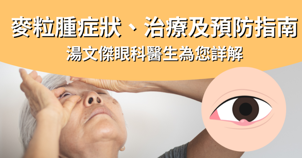
精選圖片_麥粒腫 : 原因、症狀、治療及預防指南

Corns: Causes, Symptoms, Treatment, and Prevention Guide

Corns of the eye, also known as eye pickers, although they are not a serious eye disease, the sudden appearance of a small lump on the eyelid can cause some inconvenience in daily life. Although this condition is common and in most cases does not require medical intervention, understanding its causes, symptoms, treatment methods, and preventive measures is essential to reduce the incidence and accelerate recovery.

This article will comprehensively introduce the relevant information about blepharitis, allowing you to better understand and manage this eye problem.

What is granuloma?

Corns of the eyelid, also known as "conjunctivitis" or "stye," are a common eye inflammation. The cause of cornification is mainly due to blockage of the oil glands at the edge of the eyelid, which leads to bacterial infection and inflammation of the gland or hair follicles, resulting in a small and painful abscess. Corns usually form on the inner or outer side of the eyelid, presenting with redness, pain, and localized heat sensation.

Additionally, many people mistakenly believe that cornoid cyst and chalazion are the same eye inflammation. In fact, the causes and severity of pain are different. Chalazion is a painless swelling caused by chronic inflammation of the eyelid glands, while cornoid cyst is caused by bacterial infection and is usually red, swollen, and painful. Chalazion is usually larger and less painful, while cornoid cyst is smaller but more painful.

Type of cornflower edema

There are mainly two types of cornoid tumors: external cornoid tumors and internal cornoid tumors.

  • External otitis: located at the base of the eyelashes, it causes a yellow pus abscess that usually heals quickly.
  • Inner eyelid cyst: located inside the eyelid, the inflammation is longer lasting and usually more painful than outer eyelid cyst.

Symptoms of wheat germ inflammation

  • Swollen eyelids
  • Local eyelid pain
  • Local heat sensation in the eyelids
  • Small abscesses appear at the edge or inside of the eyelids
  • Eye foreign body sensation
  • Eye tears
  • Swollen eyelids

Causes of cornflower edema

The cause of blepharitis usually results from excessive gland secretion from the meibomian glands, which leads to the formation of oil-filled particles within the eyelids. When these particles block the hair follicles or glands, bacteria can multiply and cause infection, leading to inflammation within the eyelids. Additionally, poor eye hygiene practices, touching the eyes, using contaminated cosmetics, and wearing contact lenses can also increase the risk of blepharitis.

Treatment methods for cornflower edema

Orbital inflammation is not a serious eye condition. In the early stages of blepharitis, you can self-treat with heat packs and massage at home to help remove the oil from the inner eyelids. This method should usually resolve the blepharitis within a short time. If heat packs and massage do not improve the condition, seek the help of an ophthalmologist to treat the blepharitis. Treatment options include using blepharitis ointment, oral antibiotics, and surgery.

  1. Heat compressApply 4-5 times daily for about 10-15 minutes each time, helping the abscess to drain out on its own.
  2. MassageUse your fingers to gently push. If the eyelash is on the upper eyelid, push the fingers down from the eyelid; if the eyelash is on the lower eyelid, push them up from the eye bag.
  3. Macular edema ointmentUse eyelid inflammation ointment or eye drops under doctor's guidance to control the infection.
  4. Oral antibioticsIn cases of severe infection or complications, doctors may prescribe oral antibiotics.
  5. Surgical drainageFor cystic acne that cannot be removed on its own, doctors may perform minor surgery to drain the pus.

How to prevent the formation of cornflower edema

  1. Keep your eyes cleanRegularly clean the eyelids to prevent bacteria from growing.
  2. Avoid touching the eyesBefore touching your eyes, wash your hands to reduce the risk of bacterial infection.
  3. Do not share towels or cosmetics with othersReduces the risk of cross-infection.
  4. Replacing cosmeticsChange your eye makeup regularly, especially the mascara and eyeliner.
  5. Using contact lenses correctlyWhen wearing contact lenses, follow hygiene guidelines to avoid bacterial infections.
  6. Managing skin healthTreating skin diseases such as acne and seborrheic dermatitis reduces the risk of ingrown eyelashes.

By understanding the causes, symptoms, and treatment methods of conjunctivitis, we can better prevent and manage this common eye problem.

If you experience persistent or severe symptoms, please seek medical attention immediately to receive professional treatment recommendations. If you have any further questions, we recommend that you contact your doctor at any time.The ophthalmologist conducted an examination.。

Common problems

Is a corn wart the same as a papule?

No, granuloma is a painful abscess caused by a bacterial infection, while a sebaceous cyst is a painless lump caused by gland blockage.

Will the pimple disappear on its own?

Yes, many cases of follicular lymphoma resolve on their own within a few days to a week, but proper treatment can speed up the recovery process.

How long does a corn cyst usually take to disappear?

It usually takes one to two weeks, but if the infection is severe or not treated properly, it may take longer.

Can you squeeze a pimple?

It is not recommended to squeeze the pus-filled cyst; doing so may worsen the infection and cause complications.

Do I need to see a doctor for granuloma?

If the granuloma does not subside, the pain worsens, or it affects vision, you should seek medical attention immediately.

What can't you eat with cornflower edema?

There are no specific dietary restrictions, but maintaining a balanced diet and good nutrition can help improve immunity.

Does cornflower edema have a contagious nature?

Corns are not contagious in themselves, but the bacteria that cause infection can spread, so good personal hygiene should be maintained.

Can you puncture a corn blister?

It is not recommended to puncture the corn itself; doing so may lead to serious infections such as cellulitis and inflammation in the brain; it is recommended to have it treated by a doctor.

Corns: Causes, Symptoms, Treatment, and Prevention Guide Read More »

黃斑病變食療

Treatment for macular degeneration: 6 essential nutrients at a glance

Macular degenerationMacular degeneration often occurs in people over 65, hence it is also known as age-related macular degeneration (AMD). People who suffer from this eye condition experience blurred central vision and distorted images, and in severe cases it can even lead to blindness.

Since macular degeneration is an incurable eye disease, once it is detected, only certain treatment methods or dietary supplements can slow down the progression of macular degeneration. Therefore, preventing macular degeneration has become very important, and changing certain dietary habits or eating patterns to prevent macular degeneration is one of the most direct methods. This article will introduce the important nutrients and dietary habits for macular degeneration treatment to help everyone maintain eye health.

6 Major Nutrients for Preventing Macular Degeneration

The following are reference values for nutrients and some corresponding foods that are helpful in preventing macular degeneration:

  • Lutein: Spinach, asparagus, carrots, cauliflower, pumpkin, etc.
  • Vitamin A: Red sweet potatoes, egg yolk, beef liver, mango, goji berries, etc.
  • B vitamins: Salmon, pork, beef tallow, milk, nuts, etc.
  • Vitamin C: Lemons, Guavas, Oranges, Papayas, Strawberries, etc.
  • Vitamin E: Salmon, almonds, cashew nuts, wheat germ, olive oil, etc.
  • Zinc: Whole grains, seafood, meat and beans

Flavonoids

Beta-carotene is an important substance that forms the retina and the macular region. It is the main pigment source of the retina. It not only has antioxidant effects but also absorbs harmful light, reducing the damage to the eyes and reducing the risk of macular degeneration.

Vitamin A

Adequate supplementation of vitamin A can maintain the normal function of retinal epithelial cells and help promote the metabolism of the cornea and retina.

Vitamin B group

Vitamin B complex includes various vitamins such as B1, B2, B6, and B12, which effectively maintain visual acuity and retinal nerve cells and prevent cell aging. These vitamins help promote blood circulation in the eyes, eliminate eye fatigue, and are recommended for people with excessive eye strain to supplement these nutrients regularly to prevent macular degeneration.

Vitamin C

In addition to helping prevent macular degeneration, supplementing with vitamin C can also prevent eye diseases such as cataracts, glaucoma, and retinal degeneration. Vitamin C possesses extremely powerful antioxidant properties that protect the eyes from free radical damage, and also promotes blood circulation in the microvessels of the retina, thereby preventing macular degeneration.

Vitamin E

Vitamin E has the ability to remove free radicals and also slows down the oxidation of lutein, maintaining eye health and preventing macular degeneration. In addition, vitamin E is mainly concentrated in the retinal pigment epithelial cells and photoreceptor cells, making it one of the essential nutrients for maintaining retinal health.

鋅

Zinc itself possesses considerable antioxidant capacity, and has been confirmed by many scholars to effectively maintain eye health and prevent macular degeneration. Additionally, zinc in the eye helps vitamin A function in metabolism, maintaining normal retinal epithelial function and maintaining eye health.

Dietary habits to prevent macular degeneration

In addition to specific nutrients, good dietary habits are also key to preventing macular degeneration. Here are some dietary practices that can help improve vision health:

Low-glycemic diet

People with diabetes are more likely to develop macular degeneration or a range of eye diseases than the general population. This is because high blood sugar levels can damage the retina and even cause it to detach. Therefore, ophthalmologists generally recommend diabetics follow a low glycemic diet to reduce glycation end products and control blood sugar levels while also preventing eye diseases such as macular degeneration and retinal detachment.

A low-glycemic diet, as the name suggests, is one that:Eat low-carbohydrate foods to reduce fluctuations in blood sugar levels, such as whole grains, vegetables, and lean meat.Both of these are low-glycemic foods; while white rice, white bread, and processed meats are high-glycemic foods.

In addition to people with diabetes, ordinary people can also consider adopting a long-term low-glycemic index diet to maintain their health. The simplest way to implement a low-glycemic index diet is to follow the diabetic diet, which not only prevents macular degeneration but also prevents the onset of diabetes.

Mediterranean diet

A study in Australia found that participants who followed the Mediterranean diet were 41 percent less likely to develop macular degeneration than those who did not follow the Mediterranean diet, confirming that the Mediterranean diet can prevent macular degeneration.

Mediterranean dietConsists primarily of fresh vegetables, fruits, fish, olive oil, and nuts, with a small amount of red meat and dairy products.This diet is rich in antioxidants and unsaturated fatty acids, which can effectively reduce the inflammatory response in the eyes and help prevent macular degeneration.

If you want to prevent or improve macular degeneration through a Mediterranean diet, it is recommended to consult an ophthalmologist for further advice or to get more knowledge and recipes to prevent macular degeneration from your nutritionist.

Balanced diet

If you are unable to follow the low-glycemic diet or Mediterranean diet for preventing macular degeneration due to work or other reasons, then try to ensure a balanced daily intake of nutrients as much as possible.Eat two servings of fruit and five different colors of vegetables dailyReduce your intake of fast food or high-fat, high-salt, and high-sugar foods. If necessary, you can also have a nutritionist create a preventive eye disease treatment plan for you or take appropriate supplements. Maintaining a balanced diet not only helps prevent macular degeneration but also maintains good health.

If you suspect you have macular degeneration, do not blindly rely on folk remedies or alternative treatments; it is essential to consult an ophthalmologist as soon as possible and undergo a detailed examination so that a professional doctor can develop the most effective treatment plan for you.Treatment plan。

Common problems

What are some recommended remedies for macular degeneration?

If you only want to prevent macular degeneration, you can simply drink any soup containing the appropriate amount of various nutrients; for patients who have already been diagnosed with macular degeneration, you can consult a nutritionist or ophthalmologist to choose a soup that is effective in treating macular degeneration.

Will eating alone cure macular degeneration?

As of current medical knowledge, macular degeneration has no cure, but proper treatment and dietary management can slow the progression of macular eye disease. It is recommended that if you suspect macular degeneration, you should seek immediate medical attention.

Treatment for macular degeneration: 6 essential nutrients at a glance Read More »

飛蚊症原因及治療方法

Causes, Symptoms, and Treatment Methods of Glaucoma: An Eye Specialist Explains in Detail

Floaters are a common vision problem in which patients see floating black dots, jellyfish-like shapes, and threadlike structures in their field of vision. There are many different types of floaters, and each patient’s description of the shape is unique. Is it possible to cure floaters? Will floaters resolve on their own? Dr. Tong Weijian, an ophthalmologist, will answer these questions for you.

What is fly eye syndrome?

Floaters are medically known as vitreous degeneration. There is a transparent gel-like substance called the vitreous in the eye, located in the center of the eye, behind the crystalline lens, and before the retina. In normal conditions, the vitreous fills the entire vitreous cavity, supporting the shape of the eye and allowing light to be refracted smoothly onto the retina, thereby forming an image. If the vitreous degenerates, the gel-like substance within the vitreous will undergo liquefaction, forming tiny fibers that float within the vitreous cavity, which is what patients see as black spots.

What causes fly eye syndrome?

The main causes of fly-eye can be roughly divided into three types: physiological, degenerative, and pathological. If there is any suspicion, patients are advised to seek immediate medical attention from an ophthalmologist.

Rationality

Retinal vein occlusion is common in people under 40 years of age or those who spend long hours looking at screens, such as car painters, teachers, and those who work at computers for long periods of time. In general, up to 80% of cases of retinal vein occlusion are considered physiological, and patients may feel black dots in their eyes or see lines; these are impurities in the vitreous humor. There are currently no medications or surgeries to treat this type of retinal vein occlusion, but there is no need to worry; these black dots will disappear over time within the field of view.

Degradation

There is a transparent gel-like substance called the vitreous body inside the eye, located behind the lens and before the retina. As we age, this vitreous body deteriorates and flakes off, forming tiny fibers that float inside the vitreous body, causing us to see blurry images.There are black spots on the eyes.Or it may be a black spot; for some people, however, they see a series of insect-like objects. As long as the black spots in the eye do not become more numerous or remain relatively fixed in position, it is considered a benign form of fly-eye and there is no need to be overly anxious.

Pathological

The cause of pathological floaters is due to other eye problems or systemic vascular diseases, such as external force pulling or impact, or systemic vascular diseases such as eye floaters caused by wind in the eyes, diabetes, and high blood pressure. These can all lead to vitreous detachment and bleeding, with black spots or even black patches in the eyes. If this type of floaters is not treated aggressively, it can lead to permanent blindness. Patients are advised to seek treatment for improving floaters as soon as possible.

What are the symptoms of fly eye syndrome?

The early symptoms of Glaucoma are easy to recognize. Patients may feel a floating object in their eyes, which they can see but cannot grasp. This is especially noticeable in situations with strong light or a bright background. There are many different visual forms of Glaucoma, so the shape seen by each patient is also different:

If you experience worsening symptoms of fly eye syndrome, such as a dramatic increase in the number of flies, flashing lights in the eyes, dark shadows in the eyes, blocked vision, or blurred vision, you should immediately seek medical advice from an ophthalmologist for a detailed examination and to receive treatment from a doctor to improve the symptoms of fly eye syndrome.

Treatment methods for fly eye syndrome

In the early stages of fly-eye, no immediate treatment is necessary. Most people adjust to the presence of the dark spots after a period of time. Over time, these dark spots may settle at the bottom of the vitreous humor and no longer affect vision. However, if you notice more dark spots, black shadows in your eyes, or feel that these dark spots are affecting your daily life, you may consider laser treatment or a fly-eye surgery to improve the dark spots in your eyes.

Laser therapy

Laser treatment is suitable for larger and concentrated vitreous fibers. By shattering these fibers into smaller fragments with a laser, it can improve and eliminate the symptoms of fly-eye. Laser treatment is performed under local anesthesia; there is no pain during the procedure, and the entire treatment takes about 15 to 20 minutes. However, not everyone is suitable for this treatment; it is not advisable if the fibers are loose or located too close to the retina or lens, as this could cause scarring.CataractOr secondary complications such as macular damage.

Corneal transplant surgery

Glaucoma surgery involves removing the vitreous humor from the eye through a small incision and replacing it with a solution to maintain the eye’s shape. The procedure takes about 10 to 15 minutes. Although this procedure can effectively remove the vitreous humor, it can also lead to bleeding or retinal tearing, which in turn forms new floaters. Therefore, most doctors do not recommend using this procedure to treat glaucoma.

Additionally, many patients try to improve their eye degeneration symptoms by taking eye drops. They ask ophthalmologists to recommend eye drops for glaucoma. It is important to note that eye drops cannot penetrate the vitreous humor, so simply using eye drops alone cannot treat vitreous degeneration. Dr. Tu reminds us to never blindly follow the recommendations of non-professional eye doctors for glaucoma.

Prevention methods for fly eye disease

Migraine is mostly caused by age increase andHigh myopiaThis leads to degeneration of the vitreous humor. Currently, there is no complete solution to prevent the development of fly-eye syndrome. However, we can slow down the symptoms of eye degeneration through daily care. Here are some ways to prevent the development of fly-eye syndrome:

  • Avoid excessive use of electronic devices (use them for 1 hour, take a 5-10 minute break).
  • Read or work under as much natural light as possible to avoid worsening myopia.
  • Ensure a balanced daily diet and reduce the intake of foods high in oil, salt, and sugar.
  • Eat a variety of foods containing various nutrients such as carotenoids, anthocyanins, and vitamins.
  • Regularly undergo physical examinations and eye examinations

Common problems

How to eliminate fly eye syndrome?

If the black spots in front of your eyes are causing significant disruption to your daily life, you may consider laser treatment or vitreous removal surgery to eliminate the floaters.

Why do I suddenly develop fly eye syndrome?

The occurrence of fly-eye may be caused by physiological, degenerative, or pathological factors. It is recommended that you consult a qualified ophthalmologist to accurately determine the cause and treatment method of your fly-eye condition.

Will fly eye disease disappear on its own?

Migraine headaches do not go away on their own, but most people gradually adjust to their presence after a period of time.

What will happen if I ignore the fly eye?

Benign floaters generally do not require treatment, but it is important to note whether the spots become more numerous. If you feel that your field of vision is severely obstructed, you must seek medical attention immediately to avoid delaying treatment and causing blindness.

Is blepharitis a side effect of cataract surgery?

The complications of hernia surgery do not include fly eye syndrome. Some patients have this condition because of other conditions.After undergoing cataract surgeryPeople often discover they have a retinal detachment after the surgery because their vision was too blurry before the surgery, which meant they didn't realize they had the condition in the first place.

Causes, Symptoms, and Treatment Methods of Glaucoma: An Eye Specialist Explains in Detail Read More »