遠視和老花_封面圖

遠視和老花有甚麼分別?詳解成因及治療方法

老花和遠視都是在看遠距離事物時視野清晰,看近時感覺模糊,因此人們常常混淆老花和遠視。然而,老花和近視是兩種完全不同的視力問題,各自的矯視方法也有些許不同。下文將爲你𨤳清老花和遠視的分別,及如何矯正老花和遠視。

What is presbyopia?

老花是一種與年齡相關的視力問題,當人們達到40歲左右時,眼睛的水晶體會逐漸失去彈性,使得眼睛在看近距離的物體時變得困難,尤其是在閱讀小字體或進行近距離工作時,常常需要把它們拿遠離才看得清。不但如此,常時間用眼還會容易導致頭痛、眼睛酸痛等等症狀。

老花是大家都無法避免的視力問題,假設你沒有近視、遠視或其他視力問題的話,一般人會在40歲開始出現100度左右的老花眼,其後每年的老花度數會平均增加10度;直到60歲後,度數就會逐漸趨緩。


以下老花度數表只是一個大致參考,老花度數一般以英文字母“D”(Diopter,度)來衡量,例如+1.00D,就是老花度數為100度。

Comparison table of age and presbyopia
agePresbyopia degree
40歲+1.00D
50歲+2.00D
60歲+3.00D
After the age of 60The power slows down

什麼是遠視?

遠視一種與眼球結構有關的視力問題,一般遠視都是先天性的。如果父母其中一人有遠視的話,孩子也很大幾率患有遠視問題。

遠視主要成因是因爲眼球軸長度過短或角膜曲率太平,導致焦點落在視網膜後面,而不是直接將物體聚焦在視網膜上。與老花眼相同,遠視的人可以看清楚遠處的事物,但看近距離物體時可能會感到模糊,而且長期近距離用眼時會有頭痛或眼睛緊綳的症狀。

如何分清老花及遠視?

老花及遠視徵狀看似相同,但它們卻是兩種不同的視力問題。遠視是因爲眼球軸較短而導致事物無法聚焦在視網膜上;老花則是與年齡相關的視力問題,主要形成原因是因爲睫狀肌和水晶體功能退化,導致聚焦變得困難。

以下圖表簡單概括老花和遠視的徵狀、成因、如何預防和治療方法。

Presbyopiafarsightedness
徵狀近距離影像模糊近距離影像模糊
成因睫狀肌老化,晶狀體僵硬眼球軸較短角膜弧度平坦
高危族群年齡40歲以上Family genetics
預防方法*老花無法避免,只能減緩老花速度
避免過度用眼健康飲食40歲後定期進行眼睛檢查
*遠視是先天的,只能減緩度數加深
定期進行眼睛檢查儘早矯視
治療方法老花眼鏡老花隱形眼鏡老花矯視手術眼鏡隱形眼鏡矯正手術

有遠視還會有老花嗎?

這是因爲老花與年齡相關,人人都無法避免。但遠視的人比起近視的人會更早出現老花現象,因爲老花和遠視的度數不能互相抵消。此外,若遠視患者沒有及時矯視的話,睫狀肌可能會因爲長期過度使用而加速老化。因此,即便有遠視,隨著年齡增長依然會出現老花眼的症狀。

如何治療老花及遠視

隨著醫學技術不斷發展,矯正老花及遠視不在局限於佩戴傳統眼鏡,無論是手術或佩戴隱形眼鏡,都能做到客制化的矯視效果,同時改善老花及遠視問題。

老花眼鏡

佩戴老花眼鏡是改善老花眼的最常見方法之一。度數匹配的老花眼鏡可以在近距離提供清晰的視野,幫助患者克服閱讀和近距離工作時的困難。然而,有些人爲了方便而自行在街邊隨意購買老花眼鏡,長期佩戴下來反而會傷害眼鏡。因此,一定要尋求專業眼科醫生驗配適合你度數的老花眼鏡才是明智的選擇。

對於原本就有遠視問題的人,就可能需要再配副老花眼鏡,並且需要在不同情況下更換眼鏡,這可能會造成部分人的困擾,這時就可以考慮選擇多焦鏡片以解決同時存在的老花和遠視問題。

老花con

雖説是老花con,但其設計原理和傳統的多焦鏡片相似,因此現今的老花con不再只專注于改善老花,亦能改善遠視或其他視力問題。通常這類老花con需要找專業眼科醫生爲你驗配,也並非每個人適合佩戴隱形眼鏡,因此在驗配前需要先檢查眼睛狀況。

老花矯視手術

現代老花手術是一種根本解決老花及遠視問題的方法,讓患者不再依賴老花眼鏡或隱形眼鏡。特別是最新的Presbymax老花矯視技術,能夠同時矯正遠視,使患者能夠擁有清晰的視野,不再受到老花和遠視的困擾。

Presbymax 最大的優勢在於它可以實現遠、中、近全方為的立體視覺,無論是否有散光、近視或遠視,Presbymax老花矯視手術都能完美的解決這些視力問題。此外,這類手術時間短,,傷口小,多數患者在24小時内就能恢復日常活動,成功率極高。

對於一些不想佩戴傳統眼鏡或不適合佩戴老花con的人來説,Presbymax老花矯視手術可説是最高性價比的選擇。

遠視和老花有甚麼分別?詳解成因及治療方法 Read More »

老花和散光_封面圖

老花、散光傻傻分不清?一文睇清

隨著年齡的增長,許多人可能會開始發現看事物變得模糊,閲讀變得困難,這種情況通常稱爲老花。然而,罹患散光眼的患者在看事物時同樣也會遇到視力模糊的問題,因此,很多人常常混淆老花和散光。這兩者該如何區別?老花和散光同時出現的話又該如何處理?下文將一一為你解釋。

What is presbyopia?

老花是一種隨著年齡增長而發生的視力問題。老花主要影響到人們對近距離物體的清晰度,尤其是在閱讀小字或執行細微工作時。

老花發生的主要成因是因爲眼睛中的晶狀體變得僵硬,使其難以調整焦距以看清近距離的物體。這種變化是由於眼睛組織的老化,導致晶狀體失去彈性和靈活性。老花通常在40歲左右開始出現,並會隨著年齡的增長而加劇,直到60歲後度數的上升趨勢才會逐漸減慢。

什麼是散光?

散光是一種屈光不正的現象,可分爲先天性和後天造成,無論是哪種散光,其主要症狀都是看事物時會出現曡影影像模糊或扭曲的情況。

在正常的情況底下,光線會通過角膜、水晶體和玻璃體後,在視網膜上聚為一個焦點,形成清晰的影像。如果角膜的弧度不規則的話(即,角膜不是呈正圓的半球體),光線就無法正常通過,形成多焦點的現象。

老花和散光的分别

老花和散光是兩種不同的視力問題。散光是指光線聚焦不同,導致遠近距離畫面出現模糊或扭曲;而老花則是因爲眼球結構老化造成近距離視力喪失。

以下圖表簡單概括老花和散光的徵狀、成因、如何預防和治療方法。

Presbyopia散光
徵狀近距離影像模糊閲讀困難遠近事物有曡影影像扭曲
成因睫狀肌老化,晶狀體僵硬角膜弧度不規則
預防方法避免過度用眼健康飲食40歲後定期進行眼睛檢查*老花無法避免,只能減緩老花速度良好的燈光下閲讀避免長時間使用電子產品定期進行眼睛檢查*先天性散光無法避免,只能維持良好用眼習慣,避免散光加速惡化
治療方法老花眼鏡老花隱形眼鏡老花矯視手術散光眼鏡散光隱形眼鏡手術

有散光會有老花嗎?

老花是一種眼睛老化的現象,每個人都會有老花眼,只是來的快或慢而已。因此,無論是否有近視、遠視或散光問題都會患上老花。

如何同時治療老花眼和散光?

上文提到,老花和散光是兩種完全不同的視力問題,但現有的老花矯視方法一般都能同時改善散光。一般輕微的散光(100度以内)並不需要特別治療,同時擁有老花眼和散光的患者可以在需要時佩戴老花眼鏡或老花con,如果感覺到視力不佳或感覺影像怪怪的,則需要向專業眼科醫生另尋方案。

以下是一些能夠同時改善老花和散光的普遍治療方案:

漸進式眼鏡

漸進式眼鏡能夠同時改善老花、散光、近視、遠視等問題,增加影像的清晰度。然而,由於特殊的鏡片設計,初次佩戴時可能會感到不適,需要一段時間來適應。對於一些人來說,佩戴眼鏡可能在工作上造成不便或覺得不美觀,因此可以考慮選擇佩戴隱形眼鏡,同樣能夠有效改善老花和散光問題。

漸進式隱形眼鏡

漸進式隱形眼鏡的原理和漸進式眼鏡相同,能同時改善多種視力問題。但是如果沒有佩戴過隱形眼鏡的人可能需要時間去適應它,同時也需要根據醫生指示使用,以眼部避免感染。然而,這類con的最高散光度數只能在大約在225度左右,若你的散光度數太高的話,就只能尋求眼科醫生擬定其它矯視方案。

角膜塑型片(Ortho-K)

角膜塑型片是一種矯正型隱形眼鏡。散光眼患者在睡覺時佩戴可以調整角膜弧度,睡醒后取下便能擁有正常的角膜弧度,這時患者就只需要佩戴老花眼鏡便能看清楚近距離事物。

Orthopaedic surgery

隨著醫療科技不斷地進步,現今的矯視手術已有多種選擇。對於散光和老花度數較深的患者,可以參考眼科醫生的建議,選擇合適的矯視手術方案,以重拾視力。

然而,要注意的事,老花會隨著年齡而加深,因此,在矯視手術的幾年後仍可能需要佩戴老花眼鏡或老花con來保持良好的視覺。

無論選擇哪種治療方法,都應該根據個人的情況和需求來進行選擇。因此,在決定治療方法之前,最好向眼科醫生諮詢以獲得專業建議。

老花、散光傻傻分不清?一文睇清 Read More »

How to choose the prescription of reading glasses?_Cover picture

How to improve the degree of presbyopia?

Presbyopia is an age-related phenomenon of natural aging that is caused by the deterioration of the ciliary muscle of the eye and the aging of the lens. Presbyopia can make it difficult to focus on close objects, resulting in blurry images, especially when reading with your phone or newspaper away from you. In addition, the presbyopia will continue to deteriorate with age, so if you want to have clear vision, you must wear reading glasses or seek other vision correction solutions from your doctor.

How to identify presbyopia?

Presbyopia usually begins around the age of 40 and can continue to worsen with age. Here are some early presbyopia symptoms:

  • When reading, you need to zoom in or hold the text a little farther away to see it clearly
  • It is difficult to focus when reading, and it is easy to feel headaches
  • Eyes tend to feel tired and sore
  • When looking at near objects immediately after looking at a distance, the focus will slow down
  • People who are nearsighted begin to feel that they can see clearly without glasses
  • It's hard to see in dimly lit places

Although there is currently no cure for presbyopia, there are several options to correct vision, such as wearing reading glasses, presbyopia, laser correction or surgery. If you notice that you are starting to experience any of the above symptoms, it is advisable to seek an eye doctor for an examination, who will usually confirm the diagnosis through an eye test and presbyopia measurement, and then recommend the appropriate correction method.

How to calculate the degree of presbyopia

The degree of presbyopia is generally measured by the English letter "D" (Diopter, degree), for example, +1.00D, that is, the degree of presbyopia is 100 degrees. Assuming you don't have myopia, farsightedness or other vision problems, the average person will start to develop presbyopia of about 100 degrees at the age of 40, and then the presbyopia will increase by an average of 10 degrees per year, and the prescription will gradually slow down after the age of 60.

Comparison table of age and presbyopia
agePresbyopia degree
40歲+1.00D
50歲+2.00D
60歲+3.00D
After the age of 60The power slows down

The above presbyopia prescription table is only a rough reference, the actual situation varies from individual to individual, it is recommended that you seek an ophthalmologist for an eye examination, and obtain the exact presbyopia prescription before buying the right reading glasses.

Can myopia and presbyopia cancel each other out?

Myopia and presbyopia are two different vision problems with different causes. Myopia is caused by a refractive error in the eye, which makes it difficult to see distant objects, while presbyopia is a deterioration of the ciliary muscles and lenses, resulting in blurred images when looking at near objects. However, people who have both myopia and presbyopia can see near things clearly without glasses at a certain age, because the degree of myopia is exactly the same as the increase in presbyopia, so they may mistakenly think that they do not have presbyopia.

Still, myopia and presbyopia do not cancel each other out, as myopia does not decrease with age. People with both myopia and presbyopia do not need glasses to see near objects, but they still need glasses to see things that are far away. Therefore, once you notice presbyopia, you need to consider changing glasses with multifocal lenses or seek other methods of vision correction from a professional ophthalmologist.

How to choose reading glasses?

Once presbyopia is detected, the most convenient solution is to wear reading glasses. Although the prescription will still deepen after wearing reading glasses, this can greatly reduce the level of eye fatigue and reduce the burden of seeing near objects.

In addition, some people may buy reading glasses on their own, but each person's reading power may be slightly different, and some people may also have myopia or farsightedness, and wearing glasses with inappropriate prescription will only cause discomfort.

In terms of lens selection, there are two options on the market: monofocal lenses and progressive lenses. Monofocal reading lenses can only see objects at a fixed distance and are only worn when you need to see near objects. For people with nearsightedness or farsightedness, it is necessary to change glasses with different functions frequently. Progressive lenses, on the other hand, allow you to see objects at far, medium and near distances at the same time, but these lenses are designed to be more specific and need to adapt to time.

If you find it inconvenient to wear traditional reading glasses, or if you are not suitable for wearing glasses for work reasons, you can also consider presbyopia progressive con. It should be noted that compared to traditional reading glasses, reading con with the eyeball needs to be used in strict accordance with the doctor's instructions, especially in the maintenance and cleaning of presbyopia, to avoid bacterial infection or other eye health problems.

Presbymax presbyopia laser vision correction surgery

In addition to wearing reading glasses or reading glasses, there is also a Presbymax presbyopia laser surgery that can solve vision problems such as presbyopia and myopia at one time, allowing you to completely abandon reading glasses.

Presbymax is indicated for presbyopia patients aged 40 to 60 years without cataracts. The doctor will use a laser to polish the cornea to change the curvature, and correct myopia, farsightedness, and astigmatism to close to 0 degrees in the main eye, so as to see long-distance images. The other eye reserves or creates about 150 to 250 degrees of myopia for viewing close-up images and counteracting presbyopia, and a slight spherical aberration is added to increase the depth of field of both eyes, extend the focus, and change the chromatic aberration, thus creating a 'fusion zone' for both eyes. This not only makes the far, medium and near distances clear, but also does not affect the original color and three-dimensional sensitivity, and there will be no glare problem.

In addition, the surgery can be completed in as fast as 10 minutes, safely and without discomfort, most people can return to normal activities within 24 hours after surgery, and perfect vision can be restored within a few weeks, which is suitable for most presbyopia patients.

Whether you choose reading glasses, presbyopia, or orthopedic surgery, you should choose according to your lifestyle and seek the advice of a medical professional.

FAQ

How many degrees is presbyopia serious?

The severity of presbyopia usually depends on the degree. Generally speaking, the higher the presbyopia degree, the more pronounced the effect. But everyone's situation is different, some people may be bothered by only a slight presbyopia, while others may need a higher number of glasses to achieve noticeable improvement.

Does the presbyopia increase?

As we age, the presbyopia usually increases gradually and does not become lighter, and if you suddenly feel that your presbyopia is getting better, it may be a sign of cataracts. Therefore, doctors generally recommend that patients with presbyopia have regular eye checks and change their glasses when necessary to cope with changes in presbyopia.

How to choose the prescription of reading glasses?

The prescription of reading glasses should be determined based on the results of your eye examination and the doctor's recommendation. An eye professional will help you measure your presbyopia and help you choose the best glasses for your needs. Remember to check regularly to make sure your prescription is still suitable for your needs.

Regardless of the degree of presbyopia, early identification and wearing the right reading glasses is essential to maintain good vision and quality of life. It is recommended that everyone must have regular eye check-ups to ensure your eye health and vision condition.

How to improve the presbyopia if the degree of presbyopia increases or does not decrease? Read More »

Cover image of an article with high myopia

High myopia threatens vision, cataract symptoms can not be ignored.

High myopia itself does not directly cause serious harm to vision, as there are a variety of ways to help improve myopia. However, the formation of high myopia is due to changes in the structure of the eyeball, so even if vision is improved using other methods, it is still necessary to face a series of complications that can lead to deep myopia, such as cataracts, retinal perforation and even peeling, glaucoma, etc.

If left untreated, these problems can lead to permanent blindness. Therefore, people with high myopia need to have an in-depth understanding of these potential risks and have regular eye exams to ensure healthy vision and deal with any issues promptly.

Definition of high myopia

High myopia is a relatively severe level of myopia. Myopia of 600 degrees or more, medically defined as high myopia. Its formation is associated with changes in the length of the eyeball.

The normal length range of an adult eyeball is 22-27mm. If the length of the eyeball is greater than 27mm, it will cause high myopia. In almost all cases, genetics play a role in the development of high myopia. If both parents are nearsighted, then their child also has a greater chance of developing high myopia.

High myopia

Complications of high myopia

High myopia itself usually does not cause vision loss. However, people with this condition are more likely to develop several other sight-threatening diseases.

Retinal perforation or even detachment

The retina is the imaging site of the eye, located in the inner layer of the eyeball wall, and is a transparent film. The retina of people with high myopia will be more fragile than that of normal people, because the retina will be stretched and thinned after the eyeball becomes longer, and the retina will be more prone to perforation or rupture after thinning, and retinal detachment may occur in severe cases.

Glaucoma

Glaucoma is an eye disease in which the optic nerve is damaged due to abnormal pressure in the eyeball due to excessive or inability to drain fluid from the eyeball. In people with high myopia, the eyeball is too long, so the optic nerve or supporting tissues and blood vessels inside the eye are under more pressure, which leads to changes in intraocular pressure and the formation of glaucoma.

Cataract

People with high myopia are more likely to develop cataracts than the general population because the structure of the eye has changed, which may increase the burden on the lens, which in turn will lead to accelerated aging of the lens and the formation of cataracts.

Macular lesions and bleeding

Because of the long axis of the eye, there may be cracks in the basement membrane of cells below the macula of the retina in people with high myopia. Due to the body's self-healing function, new blood vessels grow from the choroid to the retina. In addition to occupying the photoreceptor cell layer and affecting the central visual field, these new blood vessels are susceptible to damage and cause bleeding, blood clots, and scar tissue, which can affect central vision.

Floaters

There are two main causes of floaters caused by high myopia. In the case of high myopia, the eye will age at an accelerated rate, which can lead to changes in the structure of the fibers inside the vitreous humor, and when light enters the eye, these fibers are projected onto the retina, like a mosquito floating in the eye.

Another cause is retinal detachment or tear caused by high myopia, which leads to the appearance of floaters.

High myopia test

Ophthalmologists will recommend patients with high myopia to undergo a comprehensive eye examination once or twice a year, and once symptoms are found, they can be treated immediately, making it less difficult to treat.

In the examination of high myopia, the ophthalmologist usually performs the following tests depending on the individual's situation:

  • Visual acuity test: An eye test is the most common eye test and is used to determine if the eye power is stable.
  • Fundus examination: Fundus examination will use color fundus photography to check for lesions or abnormalities in the optic nerve, blood vessels, retina, vitreous, etc.
  • Intraocular pressure and visual field test: Intraocular pressure and visual field test can assess whether you are at risk of developing glaucoma, and if signs are found during the examination, the doctor can treat it before it affects your vision.
  • Ocular CT: Ocular CT is an optical coherence tomography (OCT) that can help detect macular lesions early.
  • Slit-lamp examination: The doctor can clearly observe the eyelids, conjunctiva, sclera, cornea, anterior chamber, iris, pupil, lens and anterior 1/3 of the vitreous through the slit-lamp microscope, and can determine the location, nature, size and depth of the lesion.

Regular eye exams can detect any underlying eye problems early and prompt appropriate treatment measures to ensure healthy vision.

Even if laser vision correction surgery reduces the degree of high myopia, complications cannot be avoided

In recent years, some patients with high myopia have undergone laser vision correction surgery, and their myopia has decreased, so they have forgotten to continue to pay attention to the problems that may be caused by high myopia. But in fact, laser correction only changes the curvature of the cornea, not the length of the eyeball. Therefore, all complications arising from the length of the eyeball will not be reduced by LAIS!

High myopia causes cataracts to deepen rapidly

In a recent study of cataracts and cataract surgical outcomes for high myopia, researchers found that cataracts in highly myopic eyes occurred more quickly compared to normal eyes. In addition, there is a higher prevalence of comorbid diseases and complications (e.g., retinal detachment) in the eye with high myopia.

Therefore, people with high myopia must have a routine eye exam so that their ophthalmologist can monitor the health of their eyes and look for these symptoms as well as signs of other complications of severe myopia.

High myopia threatens vision, and cataract symptoms cannot be ignored Read More »

Cataract Prevention Article_Cover Image

What can be done to prevent cataracts?

Cataract is an eye disease in which the previously transparent lens of the eye becomes cloudy and affects vision. Patients may feel a layer of white fog obscuring them when they look at objects, making it difficult to see things in front of them, which not only greatly affects quality of life, but also increases the risk of falls or other accidents. While modern medicine has provided an effective set of treatment options to deal with cataracts, preventing cataracts can avoid the inconvenience that patients experience with vision loss and the treatment process. Cataract prevention is an important concept that all ophthalmologists are trying to promote, rather than cure.

How to prevent cataracts

Lens opacity is usually caused by aging eyes, and for senile cataracts, we can prevent them by changing some lifestyle habits.

Avoid ultraviolet and blue light

The proteins in the lens absorb blue light from ultraviolet and visible light, so prolonged exposure to sunlight or prolonged staring at electronic devices can overwork the lens and become cloudy, increasing the risk of cataracts. Therefore, you should wear sunglasses in places where the sun is particularly strong, and at the same time pay attention to let your eyes rest properly and avoid playing with your mobile phone all the time.

Eat a healthy diet

EATING FOODS RICH IN ANTIOXIDANTS, SUCH AS FRUITS, VEGETABLES, AND NUTS, CAN HELP MAINTAIN EYE HEALTH. ADEQUATE INTAKE OF VITAMINS A, C, E, AND OTHER NUTRIENTS CAN ALSO HELP PREVENT CATARACTS. IF YOU ARE AT HIGH RISK OF CATARACTS, IT IS RECOMMENDED THAT YOU GET A DIET PLAN FROM A DIETITIAN OR OPHTHALMOLOGIST TO REDUCE THE INCIDENCE OF CATARACTS.

Control blood sugar levels

Diabetes is an important factor in the development of cataracts. Therefore, keeping blood sugar levels stable is one of the keys to cataract prevention. Do not consume too much sweets such as desserts and milk tea, and plant starches such as flour and rice, which are converted into glucose in the body, should not be consumed excessively. Diabetics are advised to always check their blood sugar regularly and follow their doctor's advice to maintain good blood sugar levels.

Quit smoking

The adverse effects of smoking on eye health have been extensively studied and proven. Smoking causes oxidation of lens cells, which in turn promotes the formation of cataracts. Experiments have shown that smoking can lead to the slow accumulation of heavy metal impurities in the lenses of the eyes, which in turn increases the risk of cataracts. Compared with non-smokers, smokers are 2~3 times more likely to suffer from cataracts. Therefore, quitting smoking is not only good for your health, but also one of the important measures to reduce the risk of cataracts.

Regular eye exams

Regular eye check-ups can help detect any eye problems early, and it is recommended to have 1~2 eye check-ups per year. Early detection of cataracts can make it easier to treat or surge, reducing its impact on vision.

Cataract prevention and eye supplemental food

In addition to the methods mentioned above, we can also slow down the aging of our eyes through some eye-tonifying foods or cataract diet regimens recommended by professional doctors. If you have vision problems or other health problems that require strict dietary control, you should consult a medical professional about eye care and other related issues.

Omega-3 fatty acids

Omega-3 fatty acids are a nutrient that is very important for eye health. They are found in fish, nuts, and vegetable oils. Studies have shown that getting enough omega-3 fatty acids can reduce the risk of cataracts.

VITAMIN E

VITAMIN E IS A POWERFUL ANTIOXIDANT THAT CAN HELP PROTECT THE EYES FROM FREE RADICAL DAMAGE. NUTS, SEEDS, VEGETABLES AND VEGETABLE OILS ARE ALL EYE-TONIC FOODS RICH IN VITAMIN E.

VITAMIN C

VITAMIN C IS AN ANTIOXIDANT RECOMMENDED BY THE AMERICAN OPTOMETRY ASSOCIATION TO HELP PROTECT THE EYES FROM OXIDATIVE DAMAGE AND MAINTAIN THE HEALTH OF THE BLOOD VESSELS IN THE EYES. EYE-TONIC FOODS RICH IN VITAMIN C INCLUDE CITRUS FRUITS, STRAWBERRIES, KIWIFRUIT, CAULIFLOWER, ETC.

Lutein and zeaxanthin

Lutein and zeaxanthin are important nutrients in the eye and can help protect the eyes from UV rays and free radical damage. Intake of eye-tonic foods rich in lutein and zeaxanthin, such as leafy vegetables and corn, can help prevent the occurrence of cataracts.

Keeping an eye eye healthy can prevent not only cataracts, but also other eye diseases, ensuring that you have healthy eyes as you age. If you are unfortunate enough to find cataract symptoms, please make an appointment as soon as possible for a detailed examination. Early detection and timely treatment of cataracts can greatly reduce the difficulty of treatment and maximize vision protection.

What can be done to prevent cataracts? Read More »

Featured Image_Postoperative care of cataract symptoms

How to maintain cataract surgery?

Cataract surgery involves minimally invasive removal of the cloudy lens and implantation of an intraocular lens, which takes about 10 to 30 minutes, but post-cataract surgery care takes up to 1 to 2 months.

In addition to following the doctor's instructions for medication, follow-up visits, and maintaining eye hygiene, how should patients take care of themselves to speed up the recovery time after cataract surgery?

How long do I have to rest after cataract surgery?

Modern cataract surgery is minimally invasive, usually with a 0.2 cm wound. Therefore, exactly how long to rest after cataract surgery depends mainly on the individual's vision recovery.

In general, most patients can see clearly by the second day after cataract surgery. With careful care, vision can be fully restored within 1 week to 1 month after cataract surgery. 

However, we generally recommend that patients after cataract surgery use both eyes normally as soon as possible to speed up the recovery time after cataract surgery. Within a few hours of cataract surgery, if you can see clearly, you can do daily activities such as watching TV, using a computer, eating, and showering.

However, cataract surgery should be followed by careful care, medication as recommended by the doctor, eye cleaning, strenuous exercise and heavy lifting. When the eyes are tired, they should rest properly.

Precautions after cataract surgery

Although cataract surgery is considered a minor surgery, neglect of cataract surgery can lead to intraocular infections and even the risk of blindness. Therefore, proper cataract postoperative maintenance is crucial.

  1. Follow your doctor's instructions to clean your eyes and avoid scratching your eyes.
  2. Avoid collisions, heavy lifting, or strenuous exercise for 1 month after surgery to avoid affecting wound healing or displacement of the intraocular lens.
  3. Avoid bending over to wash your hair for 2 weeks after surgery, and avoid splashing sewage in your eyes when showering.
  4. Wear an eye mask before going to bed.
  5. Wear sunglasses when you go out to prevent light from shining on your eyes.
  6. If you have eye discomfort for a long time, such as severe pain, severe redness and swelling, and vision loss, please treat it immediately.

After-effects of cataract surgery

Cataract surgery is a safe and effective treatment, but there may still be some potential sequelae, but there is no need to worry too much, most of the sequelae of cataract surgery are mild or rare, as long as the vision can be restored with careful care or timely treatment after cataract surgery.

Here are some possible cataract surgery sequelae: 

  • Seeing flashes or shadows: It's fairly common to feel some flashes or shadows within a few hours or days after cataract surgery, but these symptoms go away in a short period of time and don't cause much concern.
  • High intraocular pressure: In some cases, it may cause an increase in intraocular pressure after surgery, which usually disappears after 24 hours. If the intraocular pressure continues to rise, then it may be a problem with residual crystal cortex, inflammation, viscoelastic residue, etc., and you need to seek medical attention immediately.
  • Light sensitivity: After surgery, patients may be more sensitive to bright light, so remember to wear sunglasses when you go out. Over time, the eyes will naturally adapt and the symptoms of light sensitivity will lessen.
  • Posterior capsule opacity: After surgery, some patients may have blurred vision due to the proliferation of epidermal cells in the posterior capsule surrounding the lens. This condition can be corrected with laser treatment, which can improve the symptoms of the eyes.
  • Retinal detachment: If you suddenly see something floating or flashing lights at some point after cataract surgery, making it difficult to see what is in front of you, it is most likely a symptom of retinal detachment, and consult an ophthalmologist immediately for further treatment.
  • Floaters: Some patients find black spots in their eyes after cataract surgery, because their vision was too blurry before cataract surgery to detect the presence of floaters. Floaters are one of the symptoms of eye aging, and if there is no sudden increase in black spots, you can ignore it.

Patients should take proper care of cataract surgery and follow up on time, and seek immediate assistance from an ophthalmologist if there are unusual visual changes after cataract surgery.

Cataract Surgery Frequently Asked Questions

1. How long should I wear sunglasses after cataract surgery?

1 to 2 weeks. After cataract surgery, patients will suddenly feel that the environment becomes brighter and that they can see things clearly, which may cause them to feel uncomfortable with luminosity. Therefore, during the 1st and 2nd weeks after surgery, we recommend that patients wear sunglasses to help their eyes adjust to the light environment.

2. How long after cataract surgery can I wash my own hair?

After the 2nd week or 1 month postoperatively. Although the wound after cataract surgery is small, there is still a risk of infection. When washing your hair yourself, sewage may splash on your eyes, so it is recommended to ask someone else to wash your hair as much as possible within the second week or one month after the surgery, and you must lie flat or on your back when washing your hair to reduce the risk of infection.

3. How Long After Cataract Surgery Can I Touch Water?

1 month. We cannot guarantee that you will not touch water for 1 month after cataract surgery, but what you can do is to avoid contact with water as much as possible. Consider using a face wipe instead of water when washing your face, while eye secretions can be cleaned with cooked water to prevent bacterial infection.

4. How Long After Cataract Surgery Can I Live a Normal Life?

After cataract surgery, it usually takes about 1 month to ensure the stability of the intraocular lens. During this time, as long as you avoid heavy lifting and strenuous exercise, you can basically return to your normal life or return to work the next day after surgery. If possible, it is advisable to take appropriate rest for the first week after surgery. However, patients still need to take good care of their eyes after cataract surgery and follow up on time.

5. How long does it take for vision to recover after cataract surgery?

Generally speaking, the vision will be significantly improved the day after cataract surgery, as long as you pay attention to maintenance after cataract surgery, your vision can be restored to 8~9% within 1 week. If the vision is completely restored and the wound is completely healed, it usually takes about 3 to 4 weeks, and the prescription will be relatively stable at this time.

 6. Will cataract recurrence after surgery?

No. However, about 5% of patients will feel that their vision will gradually become blurred after a period of surgery, because the epidermal cells proliferate in the posterior capsule, causing the posterior capsule to become cloudy, which can be improved with laser treatment. Therefore, when vision changes occur after cataract surgery, you should seek medical attention as soon as possible and treat them in time.

How to maintain cataract surgery? Read More »

Featured image_Eye sores

Picking needles in raw eyes is painful! How many patience first? Ophthalmologist analyzes how to treat eye sores and how to prevent them

Peeping into someone else's bath will cause eye sores/eye needles? How long do symptoms take to get better? Is it possible to recover by applying ointment on your own? Or do you want to see an ophthalmologist for eye sores/eye needles? I believe that many people will have similar questions, especially the most want to know, once sick, how long will eye sores / eye needles be good? Before answering these questions, we need to understand the pathogenesis.

Causes of eye sores/eye needles

Eye sores, commonly known as eye needles, are medically called "styes". Eye sores/needles usually occur near the meibomian glands and can cause redness and swelling of the eyelids. The main cause of eye sores/needles is excessive oil secretion of the meibomian glands, resulting in blockage of the glands, coupled with the lack of attention to eye hygiene on weekdays, which causes bacterial infections.

In addition, if the patient often develops eye sores/needles, it may also be because the hair follicles of the eyelashes are blocked by mites. These tiny creatures can live at the roots of eyelashes, and when they grow in large quantities, they can lead to inflammation and the accumulation of pus, eventually forming eye sores/eye picks. So in fact, it is not formed because of peeking at other people's baths.

To prevent eye sores/needles, daily eye care and cleansing are very important, especially for people who wear makeup frequently, be sure to remove makeup thoroughly and avoid sharing makeup and makeup tools with other people as much as possible. If eye sores/needles are almost intolerant and tend to occur, an ophthalmologist should be consulted to ensure that the sores/needles condition is managed appropriately.

Characteristics and symptoms of eye sores/needles

Eye sores/needles grow in different locations and correspond to different names, namely "external styes" and "internal styes". The sebaceous glands that grow on the more superficial eyelashes are external styes; The internal styes grow in the deep meibomian gland, because the pus point is on the inside of the eyelid, so the inflammation time will be longer.

At the beginning of the disease, there is only a slight foreign body sensation, and it will not be painful to the touch, and such symptoms will heal themselves as long as they rest more and apply appropriate heat. However, if there is a bacterial infection with eye sores/needles, the following symptoms will occur:

1. Symptoms of eye sores/eye needles

  • Slight redness and swelling of the eyelids
  • Minor itching and dandruff
  • There is a foreign body sensation
  • A scratching sensation when blinking

2. Severe symptoms of eye sores/eye needles

  • The eyelids are visibly red, swollen, enlarged, and even affect the field of vision
  • Pain, eyelids accompanied by slight warmth
  • The foreign body sensation is heavy
  • Transient astigmatism is present

If persistent swelling and pain are found, an ophthalmologist should be consulted immediately for eye sores/needles.

How are eye sores/needles treated?

Some patients try to pluck the affected area with a needle and squeeze the pus out when dealing with eye sores/needles, but this may cause bacterial infection, causing the wound to become inflamed or recur again, or causing cellulitis or even brain infection. Therefore, doctors do not encourage patients with eye sores/needles to be treated in this way. If you find that you have eye sores/needles, you should consult an ophthalmologist. Usually, when doctors treat and treat eye sores/needles, they will provide appropriate treatment recommendations according to the severity of the patient.

1. Mild eye sores/eye needles

To deal with mild eye sores/eye pick-and-needles, as long as you pay attention to eye hygiene, you can try to warm towels and "hot eggs" to warm the compress to make the eye sores/eye pick-needles subside on their own, the method is to boil the eggs, do not need to peel the shell, wrap them in a gauze towel, gently massage the eyes, apply 4 to 5 times a day, each time for 15 minutes, the blocked oil will melt and push out, but also reduce the blockage of the hair follicles by the oil, this method of eye sores / eye picks needles will be good without a few resistances.

However, if you find that the eye sores/needles are too painful, it is recommended to seek help from a doctor who will also prescribe some steroids or antibiotics according to the eye sores to help reduce inflammation and prevent infection.

2. Severe eye sores/eye needles

If you notice that the eye sores/needle are still inflamed and do not improve, the doctor will recommend that the person with eye sores/needle pick needle inject corticosteroids to remove the lump. However, this medication whitens the surrounding skin and is not optimal for dark-skinned people, so doctors may recommend minor surgery to remove eye sores/needles. This type of surgery is fairly simple and the wound is minimal, and there is no scarring on the appearance. If eye sores/needles still occur frequently in the same area after surgery, doctors will send the tissue to a laboratory to rule out cancer, but usually lumps on the eyelids are benign and harmless.

How good is eye sore/eye needle?

As for eye sores/eye needles, it depends on the severity of the patient. Generally speaking, mild eye sores/eye needles should be patient and wait a few days to get better, and the swelling will slowly subside; Severe patients need to be more patient, and eye sores will not improve until almost 2 weeks. If eye sores/needles are treated with resistance, they will get better in almost a few days and the course of the disease is expected to be shortened.

Whether it is a mild or severe patient, when dealing with eye sores/eye needles, the most important thing is to seek medical attention in time, and be patient to wait a few days to see if it will improve, self-treatment of eye sores/eye needles is not recommended, because this will cause more problems, resulting in the final eye sores/eye needles are almost more patient first will improve, recover, then may even take more than a whole month to recover.

Are eye sores/needles contagious?

In general, eye sores/needles are not contagious by themselves. However, if you do not treat eye sores/needles in time, which can lead to the growth of bacteria or hair follicle mites, there is a risk that it may be transmitted to the other eye or to others through contact. Sharing towels, pillows, or cosmetics with people increases the risk.

Will eye sores/eye needles be almost intolerant and often recur?

Anyone who has long sores or needles knows that the feeling is not only quite uncomfortable, but sometimes directly affects the appearance. Although this is not an emergency, it is definitely the biggest problem for some people, because eye sores/eye needles are almost intolerant and often recur. To prevent and prevent recurrence of crises, the most important thing is to start with hygiene, diet and preventive treatment.

  • Pay attention to hygiene: keep your hands clean, especially before touching your eyes; Also avoid touching your eyes with your hands and wiping your eyes with unclean towels
  • Healthy work and diet: reduce staying up late and let the eyes fully rest, in order to effectively regulate the oil secretion of the meibomian glands; Reduce the consumption of fried or spicy foods to reduce the chance of inflammation in the body

TREATMENT TO PREVENT RECURRENCE IS MORE EFFECTIVE IN PATIENTS WHO HAVE RELAPSES THAT ARE ALMOST INTOLERANT TO EYE SORES/NEEDLES, AS THEY KILL PARASITES, ARE ANTI-INFLAMMATORY, AND ANTIMICROBIAL. TAKING IPL (INTENSE PULSED LIGHT) AS AN EXAMPLE, IPL HAS PHOTOBIOREGULATORY FUNCTIONS, EFFECTIVELY STIMULATES CELL MITOCHONDRIA AND PROMOTES CELL HEALTH. TREATMENT WITH ANTIBIOTICS CONTAINING TETRACYCLINE OR MACROLIDES CAN HELP WITH ANTI-INFLAMMATORY AND BACTERIOSTATIC TREATMENT.

If not properly managed, eye sores/needles may be tolerant and often recur after a few months, or cause other more serious eye problems. Therefore, if there is eye sore/needle picking, it is best to seek an ophthalmologist to manage eye sores/eye needles.

Ophthalmologists analyze the treatment methods and prevention methods of eye sores Read More »

Featured Image_Will age-related macular degeneration get better?

Will age-related macular degeneration be okay?| Ophthalmologist says you know: senile macular disease becomes the cause and treatment method

Macular degeneration is a chronic eye disease that is often referred to as age-related macular degeneration due to blurred central vision, distorted images, and dark spots in the central area, mainly caused by the gradual aging of the eyes with age. However, some young people may develop age-related macular degeneration due to other factors. So, with current medical technology, is there a feasible solution to treat age-related macular degeneration? Will some macular degeneration be better on their own? The following is to explain the causes of age-related macular disease and surgical treatment, as well as early detection of eye problems, and early surgical treatment of age-related macular degeneration.

Type of macular degeneration

The macula is located in the middle of the retina, and most of the photoreceptor cells are concentrated in the macula, which can help us clearly see things in front of us, such as words, colors, and appearance. Clinically, macular degeneration is divided into two morphologies: dry age-related macular degeneration and wet age-related macular degeneration, of which wet age-related macular degeneration is more severe than dry age-related macular degeneration, and the treatment is also different.

Dry age-related macular degeneration

  • Dry age-related macular degeneration is the most common type, accounting for about 8~9%.
  • The cause is the accumulation of metabolites formed by the degeneration of retinal cells
  • Dry age-related macular degeneration progresses slowly and may not have a noticeable visual impact at first, but as the disease progresses, vision may gradually become blurred or lose central vision, and even develop into wet age-related macular degeneration

Wet age-related macular degeneration

  • Age-related macular degeneration is less common, and people with these macular degeneration may become blind
  • The cause is the growth of abnormal blood vessels in the macular area, resulting in the destruction of all layers of the retina, resulting in retinal hemorrhage, vitreous hemorrhage, macular edema, and retinal scarring pull
  • Age-related macular degeneration progresses more rapidly and may cause sudden blindness or severe vision loss in a short period of time

What causes age-related macular degeneration?

Senile macular degeneration is affected by a variety of factors, the most important of which is age-related, which is the main cause of central vision loss in middle-aged and elderly people over 50 years old. However, some young people may also develop macular degeneration, which may be due to congenital inheritance or acquired bad habits. Other factors include:

  • Smoking: Studies have shown that smokers are 3 to 4 times more likely to develop macular degeneration than the general population
  • Poor eating habits: Long-term intake of high-fat, high-sugar and other foods causes obesity and three highs, affects the health of eye blood vessels, and increases the risk of macular degeneration
  • LONG-TERM EXPOSURE TO INTENSE SUNLIGHT: UVA IN THE ULTRAVIOLET RAYS OF SUNLIGHT ACCELERATES THE AGING OF PHOTORECEPTOR CELLS, CAUSING DAMAGE TO THE MACULA
  • CELL GROWTH FACTOR ROLE: SOME GENETIC ABNORMALITIES IN THE HUMAN BODY OR RELATED TO MACULAR DEGENERATION, SUCH AS COMPLEMENT FACTOR B, COMPLEMENT FACTOR H, THESE GENE ABNORMALITIES HAVE BEEN SHOWN TO BE GREATLY RELATED TO THE DEVELOPMENT OF MACULAR DEGENERATION

In addition, although blue light from electronic products is rumored to be one of the causes of age-related macular degeneration, there is not enough research data to confirm this claim. However, using electronics in a darker environment may cause the macula to absorb more blue light, which can lead to various eye diseases.

What are the symptoms of age-related macular degeneration?

The symptoms of age-related macular degeneration may only occur in a single eye, but it is also possible that both eyes appear successively, if you find that the central vision is a little blurry, the image is distorted or the color becomes lighter, etc., you can basically determine that it is age-related macular degeneration, but it is still recommended to go to an eye clinic for examination to make a correct judgment.

  • Visual disturbances: Age-related macular degeneration affects central vision, and people feel that central vision gradually becomes blurred or has dark spots, making it difficult to recognize details, read, drive, and distinguish faces
  • Altered visual field: patients may feel things deformed in the central field of view, for example, straight lines may appear curved or distorted; Distance and altitude are often misjudged
  • Abnormal color vision: People with age-related macular degeneration may have problems with color perception, such as dull colors or difficulty distinguishing similar colors

MACULAR DEGENERATION SELF-TEST METHOD - AMSLER GRID

AMSLER GRID IS A QUICK WAY TO DETECT AGE-RELATED MACULAR DEGENERATION AT HOME, AND IF YOU FEEL YOU ARE AT RISK OF AGE-RELATED MACULAR DEGENERATION, YOU CAN FOLLOW THESE STEPS:

  1. Find a bright place with your eyes about 30cm away from the checkered table
  2. Cover one of the eyes with your hand and stare at the black dot in the center
  3. Repeat the above action with the other eye

If you notice that the straight line is curved, some areas are blurred or missing, or it may be a warning sign of age-related macular degeneration, you should seek advice from your ophthalmologist as soon as possible.

However, although age-related macular degeneration causes a decrease in central vision, it does not affect peripheral vision, and the risk of complete blindness is relatively low. Although age-related macular degeneration does not cause complete blindness, loss of central vision can have a considerable impact on activities of daily living. Once age-related macular degeneration is suspected, you should seek immediate consultation with an ophthalmologist for early treatment to control the condition of age-related macular degeneration.

What are the surgical treatments for age-related macular degeneration?

There are laser surgery and intraocular injections for the treatment of age-related macular degeneration, and the cost of surgery for both types of macular degeneration is also very valuable. At present, intraocular injection is the most mainstream method of treating age-related macular degeneration, and for some more serious patients, doctors will recommend surgery to treat age-related macular degeneration.

In addition, most of the treatment options for age-related macular degeneration focus on more severe wet age-related macular degeneration, and most doctors will recommend that patients consume more eye-friendly foods or supplements on a daily basis, change bad habits such as smoking, and get regular check-ups.

Laser light coagulation

In addition to the treatment of age-related macular degeneration, laser photocoagulation surgery can also treat other eye diseases such as cataracts, and its principle is to use the heat energy of the laser to destroy the abnormal blood vessels, so as to reduce blood vessel bleeding and prevent vision loss. However, this age-related macular degeneration treatment may destroy healthy tissue, and if the blood vessels are located fairly close to the center of the macula, it is advisable to find another treatment option.

Photodynamic therapy

Photodynamic therapy is a combination of drugs and lasers to treat age-related macular degeneration, in which the ophthalmologist will first inject photosensitive drops into the patient's arm, which will follow the body's blood flow and attach to the abnormally proliferated blood vessels, and then use cold lasers to stimulate the photosensitive drops, which will destroy the abnormal blood vessels, thereby achieving the effect of treating age-related macular degeneration. Unlike laser photocoagulation, this type of macular degeneration treatment surgery does not harm the surrounding tissues and is also beneficial for improving vision problems.

Anti-vascular endothelial growth factor vitreous injection

Injections in the eye are the most commonly used methods for the treatment of age-related macular degeneration, the ophthalmologist will administer an anesthetic injection on the surface of the eyeball, and then inject anti-vascular endothelial growth factor to prevent abnormal vascular hyperplasia, thereby reducing the risk of neovascular bleeding, this kind of injection treatment can not only control the condition of macular degeneration, but also improve the damaged vision, injection is quite effective in the treatment of wet age-related macular degeneration.

Another intraocular injection for age-related macular degeneration is steroid injection, which can also effectively reduce the damage of the retinal barrier by intraocular blood vessels and improve macular edema. However, it is important to note that steroids have the potential to cause other eye problems such as increased eye pressure, cataracts, etc.

New technology for the treatment of advanced age-related macular degeneration

When age-related macular degeneration cannot be treated with lasers or injections, ophthalmologists often recommend some assistive methods to improve vision, such as bifocal additional intraocular lenses in the eye or low vision glasses. Although this method may be more expensive than macular surgery, it is relatively straightforward and quick.

Low vision glasses

Smart low vision glasses project images of their surroundings in real time to the healthiest areas of the retina of low vision people and adjust them to the patient's perception. These glasses improve the patient's perception of real-world scenes by supplementing and correcting visual details. During the setup process, the ophthalmologist can intuitively adjust the glasses to help the patient find the best vision.

Bifocal additional intraocular lens

SML (Scharioth Macular Lens) is an implantable lens that can help patients "magnify" their field of view, with a specially designed central optical area that provides a high additional optical focal point of +10.0D to improve advanced vision without compromising distance vision.

Will some macular degeneration get better after surgery?

Some patients ask, "Will macular degeneration be better after surgery?" Or "Will the macular degeneration get better on its own?"

Unfortunately, age-related macular degeneration is an eye disease that cannot heal naturally, and surgery or other age-related macular degeneration treatment options can only slightly improve vision and prevent it from deteriorating further. The key is prevention, usually should take care of eye health, reduce direct eyes, supplement more nutrients such as lutein, try to delay the speed of eye aging, in order to reduce the chance of suffering from age-related macular degeneration.

After completing any macular surgery, be sure to follow your doctor's instructions to order or take your medication on time. But is it possible that as long as you take medicine on time, the condition of macular degeneration will be completely fine? In fact, patients should also pay more attention to their daily life, try to avoid rubbing their eyes, reduce eye use, and avoid smoking or construction sites and other soot-heavy environments. Macular degeneration surgery should also pay attention to diet and consume more nutrients that are beneficial to the macula, such as dark green vegetables, goji berries, egg yolks, etc. Studies have shown that daily intake of 6 mg of lutein effectively reduces the chance of macular deterioration. If the patient suffers from age-related macular degeneration due to three highs or other health problems, it is necessary to change the diet and lifestyle to control the recurrence of macular degeneration and reduce the risk of recurrence.

Will age-related macular degeneration be better? | What do ophthalmologists say: age-related macular disease becomes the cause and treatment Read More »

Myopia in children can be big or small! The control and prevention plan is clearly read in the article

Myopia is one of the most common eye problems among children nowadays. According to the Department of Health, about 18% of children aged 6 in Hong Kong have myopia. The rate of myopia in 12-year-olds is as high as 62%, which is much higher than that in other Eurasia regions. In addition to bad eye habits, if parents themselves have high myopia, the chance of children suffering from myopia will also increase significantly. Therefore, early prevention and control of myopia in children is extremely important.

Electronic products are the leading cause of myopia in children

With the popularization of electronic products, the age group of myopic children has gradually increased. Many parents use electronic products as children's pacifiers, and children develop the habit of relying on electronic products when their eyes are not fully developed, affecting the normal development of their eyes.

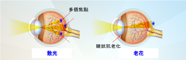
Among children with myopia, up to 80% of children myopia is caused by poor eye habits. Watching the electronic screen for a long time will make the pupil constantly adapt to the change of light source, so that the ciliary muscle that regulates the pupil is always in a state of tension, resulting in excessive eye fatigue and enhancing the risk of myopia in children. To prevent or control myopia in children, parents must strictly control the amount of time their children spend using electronic devices, encourage them to participate in outdoor activities, and reduce their reliance on electronic nipples.

Is myopia in children also true and false?

But in fact, myopia also has "true myopia" and "pseudomyopia", the former is because the eye structure has changed, so it cannot recover naturally; The latter is a manifestation of temporary eye fatigue, which can be improved as long as the eye muscles are properly relaxed. Since the two types of myopia recover in different ways, the focus should be on understanding which type of myopia children belong to.

Pseudomyopia often occurs in children under the age of 12, because the ciliary muscles of children are quite powerful, and they use electronic products for a long time in close proximity, which is prone to the inability of the ciliary muscles to relax naturally, resulting in pseudomyopia. However, as long as the eyes are sufficiently rested, children with pseudomyopia can return to normal vision.

If you suspect that the child at home has vision problems, it is recommended to seek an ophthalmologist for examination immediately, usually pseudomyopia in children can be restored, most of them are ordered to relax the eyes, so as to detect the true refractive state of the eyes, to judge true/false myopia.

Although pseudomyopia in children can recover, it is also a major sign of myopia, and if children do not correct their eye habits in time, they may also become true myopia.

How to find out if a child has myopia?

Children will not realize that they have vision problems, so parents should pay extra attention to whether children have myopia or other vision problems through observation. If the following signs of myopia occur, parents should take their child to consult an ophthalmologist:

  • Blink or rub your eyes frequently
  • Often pull the corners of your eyes or squint to see things in the distance
  • Often tilt your head to see things
  • When looking at things, the eyes are close to things

What are the consequences of neglecting to control myopia in children?

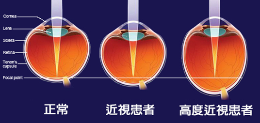
Some parents may think that if their children's myopia is not deep, they can ignore it. However, in fact, if children's myopia is not controlled, the degree of myopia will gradually increase with age, and even soar in a short period of time. If you already have myopia at a young age, it is easy to cause myopia to exceed 500 degrees.

But do you think it's just as simple as deepening myopia? No! When the degree of myopia is deeper, the chance of suffering from eye diseases such as cataracts, glaucoma, and macular degeneration will increase exponentially in the future, and severe may even lead to blindness.

  • Cataracts: People with high myopia tend to degenerate and cloudify their lenses, leading to an increased risk of cataracts.
  • Glaucoma: People with high myopia have higher intraocular pressure and are 4 times more likely to develop glaucoma.
  • Macular degeneration: Among patients with myopia of 800 degrees and myopia for 10 years, about 5~10% of patients have macular degeneration.
  • Floaters: The vitreous of highly myopic people is prone to degeneration, resulting in fibrofilamentous suspended solids.
  • Retinal detachment: The retina of highly myopic people is prone to degeneration or atrophy, and the risk of developing retinal detachment is more than 20 times that of ordinary people.

Therefore, parents should actively pay attention to the degree of myopia in children and take control and preventive measures to avoid children becoming highly myopic in the future.

What are the ways to control myopia in children?

Generally speaking, the ideal rate of myopia progression for children should be maintained at no more than 50 degrees per year. If children's myopia increases by more than 100 degrees per year, it is an unusual situation, and it may develop into high myopia in the future, and even increase the risk of complications, so the medical community has been committed to developing a variety of methods to control and improve myopia in children in recent years to slow down the progression of myopia in children.

Low-concentration atropine eye drops

Low-concentration atropine eye drops can inhibit scleral overgrowth, slow down eye elongation, and then control the progression of myopia in children, which can slow the progression by 67% in 1 year.

However, some children may experience photophobia side effects after using low-concentration atropine eye drops, or may not be clear when seeing things at close range, so caution should be taken before using low-concentration atropine eye drops.

ORTHOKERATOLOGY CONTACT LENSES (OK LENSES)

ORTHOKERATOLOGY CONTACT LENSES (OK LENSES) ARE CONTACT LENSES WORN AT NIGHT AND ARE SUITABLE FOR CHILDREN WITH MYOPIA WHOSE MYOPIA DOES NOT EXCEED 600 DEGREES. THE PRINCIPLE IS TO HELP SHAPE THE CORNEA THROUGH HIGHLY OXYGEN-PERMEABLE RIGID LENSES, ALLOWING USERS TO SEE CLEARLY DURING THE DAY WITHOUT GLASSES. STUDIES HAVE CONFIRMED THAT LONG-TERM WEARING OF OK LENSES CAN CONTROL THE DEEPENING OF MYOPIA IN CHILDREN, AND GENERALLY CAN DELAY THE SPEED OF MYOPIA IN CHILDREN TO 50%~80%.

However, orthokeratology contact lenses are only temporary vision corrections, and if they are stopped, the deepening of myopia in children will return to their original state.

IN ADDITION, THE PRICE OF OK LENSES TO CONTROL CHILDREN'S MYOPIA IS USUALLY AROUND HK$5,000~HK$10,000, PLUS THE LENSES NEED TO BE REPLACED EVERY 9 MONTHS OR SO, SO THE PRICE OF SUCH CHILDREN'S MYOPIA CONTROL LENSES MAY BE A BIT BURDENSOME FOR SOME FAMILIES.

DEFOCUSING LENSES (DIMS)

There are two degrees of defocusing lenses, namely ordinary myopia lenses in the center and peripheral lenses, which use myopic defocusing optical elements to focus peripheral images in front of the retina, thereby slowing down eye elongation and controlling the speed of myopia in children. Studies have confirmed that children who wear defocusing lenses have significantly reduced myopia by 52% within 2 years.

UNFORTUNATELY, DEFOCUSING LENSES ARE ONLY SUITABLE FOR MYOPIC CHILDREN WITH MYOPIA OF NO MORE THAN 650 DEGREES AND ASTIGMATISM OF NO MORE THAN 400 DEGREES, AND CHILDREN WHO EXCEED THIS DEGREE CAN ONLY FIND OTHER CONTROL SOLUTIONS. IN ADDITION, THE PRICE OF THESE CHILDREN'S MYOPIA CONTROL LENSES IS NOT LOW, AND MOST OF THE COMMERCIALLY AVAILABLE CHILDREN'S MYOPIA CONTROL DEFOCUSING LENSES START FROM HK$4,000, WHICH DIRECTLY DISCOURAGES PARENTS WHO WANT TO USE LENSES TO CONTROL CHILDREN'S MYOPIA.

AN EARLIER METHOD TO CONTROL MYOPIA IN CHILDREN - AI MYOPIA CONTROL PROGRAM FOR CHILDREN

As mentioned above, some eye drops to control myopia in children may have side effects, and the current high price of defocusing lenses for controlling myopia in children, coupled with the so-called "prevention is better than cure", parents are beginning to realize that to control children's myopia and the chance of myopia deepening, they should not start planning after discovering that they have myopia, but should start to control the risk of myopia in children at an earlier stage.

THE CHILDREN'S AI MYOPIA CONTROL PROGRAM IS A KEY RESEARCH AND DEVELOPMENT PROJECT IN HONG KONG IN RECENT YEARS, WITH A PREDICTION ACCURACY RATE OF MORE THAN 80%. THE PURPOSE IS TO PREDICT THE DEVELOPMENT OF MYOPIA IN CHILDREN IN THE NEXT 3 YEARS IN ADVANCE, AND CUSTOMIZE INDIVIDUALIZED TREATMENT PLANS IN ADVANCE TO CONTROL THE DEEPENING OF MYOPIA OR RESTORE VISION IN CHILDREN.

THE MAIN PROCESS OF THE CHILDREN'S AI MYOPIA CONTROL PROGRAM IS AS FOLLOWS:

  1. Fill in the questionnaire on children's habits
  2. Perform various eye examinations
  3. Enter the examination data and analyze the children's myopia in the next 3 years through artificial intelligence and big data
  4. Parents and ophthalmologists can use the reports and charts to find out whether children's myopia development is at high risk
  5. Based on the evaluation results, the ophthalmologist will customize personalized treatment for the child to delay the progression of myopia in children and reduce the risk of high myopia.

At present, the children's AI myopia control project led by ophthalmologist Dr. Tang Wenjie has been verified in 129,242 adolescent populations, with a prediction accuracy rate of more than 80%, providing early warning of myopia among adolescents.

PARENTS CAN ACCURATELY PREDICT THEIR CHILDREN'S FUTURE MYOPIA CHANGES THROUGH THE "AI INTELLIGENT PLATFORM FOR MYOPIA CONTROL IN CHILDREN", AND THE PREDICTION RESULTS CAN BE INTELLIGENTLY ANALYZED THROUGH THE BIG DATA OF THIS AI PLATFORM, SO THAT PARENTS CAN OBSERVE THE CHANGES IN THE REFRACTIVE ERROR PERFORMANCE OF THEIR CHILDREN AT DIFFERENT AGES ACCORDING TO THE PREDICTION REPORT (PARENTS CAN SEE THIS AS THE "REFRACTIVE DEVELOPMENT FILE"). BASED ON THE FORECAST REPORT, THE OPHTHALMOLOGIST WILL ALSO ANALYZE THE CURRENT MYOPIA SITUATION OF THE CHILDREN FOR PARENTS IN DETAIL, COMPARE THE NORMAL REFRACTIVE PERFORMANCE OF THE SAME AGE GROUP, AND FORMULATE PERSONALIZED MYOPIA TREATMENT PLANS.

Everyday ways to improve or control myopia in children

To prevent or control myopia, in addition to seeking the help of an ophthalmologist, parents can also help myopic children recover or improve their vision from daily life.

Establish proper eye habits

Try to stipulate that children should maintain a correct sitting posture when doing homework or reading, avoid lying on their stomachs or lying down, and should also avoid them reading or playing with mobile phones in a dim environment, and control the daily use of electronic products by myopic children, and develop the good habit of resting for 5~10 minutes every 30 minutes with their eyes.

Consume eye-friendly foods

PARENTS CAN ALSO GIVE THEIR CHILDREN MORE EYE PROTECTION FOODS SUCH AS VITAMINS A, C, E, LUTEIN, ANTHOCYANINS, ETC., WHICH CAN HELP MYOPIC CHILDREN RESTORE OR IMPROVE THEIR VISION.

Outdoor activities

It is recommended that parents take children to outdoor activities every day, such as playground play, park walking, reduce their time to use electronic products, and outdoor sunlight can also stimulate the secretion of retinal dopamine, effectively control the deepening of myopia in children.

Myopia in children develops very quickly, and the slightest attention can quickly soar. Therefore, in addition to supervising children to develop good eye habits in daily life, it is recommended that parents regularly take children for eye examinations. Once abnormalities are found, appropriate control measures and treatment plans can be taken in time to ensure the health of children's vision.

Myopia in children can be big or small! Read More »

Explain Presbymax presbyopia laser vision correction surgery

Get rid of reading glasses from now on! 【Presbymax Presbymax Laser Surgery】Helps restore youthful vision

Presbyopia is a vision problem that occurs with age, and almost everyone has presbyopia, usually in people around the age of 40. Many people with presbyopia are reluctant to wear reading glasses for fear of being ridiculed for their age, which affects their daily lives. However, now that there are more and more ways to correct presbyopia, wearing reading glasses is not the only option, such as the Presbymax presbymax laser surgery highly recommended by ophthalmologist Dr. Tang Wenjie, which can not only avoid the embarrassment of wearing reading glasses, but also restore the vision of the eyes when they were young.

What is presbyopia?

The most common symptom of presbyopia is dyslexia, reading the text on the mobile phone or newspaper can become quite strenuous, and it is necessary to constantly adjust the distance to find a clearer focus. Prolonged close movement can make it easy to feel eye strain, dryness or discomfort; When working at close range in darker environments, more light is needed to help the eye focus.

The main cause of presbyopia is the degeneration of the lens and ciliary muscle in the eyeball. This degradation causes the lens to become stiff and inelastic, which in turn affects the eye's refractive ability and ultimately vision.

Although presbyopia usually occurs in people in their 40s, however, with the advancement of technology, people are increasingly relying on electronics. Staring at the screen for a long time will cause the ciliary muscle to tighten too much, causing the eyes to enter the presbyopia stage early. Therefore, protecting eye health is extremely important for modern people. It is recommended to reduce the time spent using electronic products to allow enough time for the eyes to relax to slow down the development of presbyopia.

2. Method self-test for presbyopia

Method 1: Symptom check

The following are some common symptoms related to presbyopia, if you get more than 3 of them, you may suffer from presbyopia.

  • When looking at the phone, you need to hold farther away to see clearly
  • When looking at the subject, I want to take off my glasses, but I can see more clearly without glasses
  • Feel that your home or office is darker than before
  • Often misread numbers or words
  • Eye strain is often felt
  • Shoulder and neck aches, stiffness
  • Often frowning, dizziness

Method 2: Banknote test

  1. Take out a note and place it where you can see it best
  2. Then focus on the small print on the banknote
  3. Look away from the banknote and look at an object 2 to 3 meters away for about 5 seconds
  4. Then look back at the small print and see if you can read the small print in a few seconds

Being able to focus within 1 second is normal; More than 1 second is at risk of presbyopia; More than 3 seconds can basically determine that there is presbyopia.

If you suspect that you have presbyopia, it is recommended to consult an ophthalmologist as soon as possible for professional advice and help. Your ophthalmologist will give you the need to wear reading glasses based on your vision needs or recommend Presbymax laser surgery to improve presbyopia. Early recognition and treatment of presbyopia can help improve vision and improve quality of life.

What is the difference between presbyopia and farsightedness?

Presbyopia and farsightedness are also blind, but the causes of the two are not the same. In addition, if you find that you have vision problems, it is recommended to go for a test.

  • Presbyopia is the degeneration of the eyeball

Presbyopia is an age-related vision problem in which the lens and ciliary muscle of the eye gradually deteriorate with age, making it less focused and making it difficult for the eye to see clearly in close objects and dark environments. If presbyopia has seriously affected your life, Presbymax presbyopia correction surgery can be considered. This is because Presbymax can not only treat presbyopia, but also help improve myopia, farsightedness, astigmatism and other problems, and restore the eyes to their youthful state.

  • Farsightedness is innate

Farsightedness is a vision problem related to the structure of the eyeball. It is mainly formed by short eyeballs or weakness of the ciliary muscle. If only the eyeball is short but the ciliary muscle is developed, there will basically be no problem of blurred vision; When the ciliary muscle is weak, it needs to be forced to focus on the near object. Farsightedness is innate, meaning that most children have farsightedness, but there is no need to worry too much, as the eye develops normally, the farsightedness will be relieved.

Leave the old flowers alone, be careful of physical problems!

Many patients feel that presbyopia has little impact on life and are reluctant to face up to the problem of presbyopia, thinking that they can see clearly by simply squinting, and even reading glasses are reluctant to wear. However, this behavior will cause ciliary muscle overwork, increase the burden on the eyes, cause eye soreness and discomfort, eye pain, etc., and in severe cases, it will even spread to the shoulder and neck area, resulting in shoulder and neck pain, headache, dizziness, nausea and other symptoms.

Although presbyopia is not an emergency, the degree of presbyopia will increase with age. If presbyopia at 100 degrees is diagnosed at 40 degrees, it will increase to 300 degrees at 60 degrees, and the degree of presbyopia will increase by about 10 degrees per year. If the presbyopia degree decreases abnormally, this may be a warning sign for cataracts, and patients should seek detailed examination and appropriate correction from an ophthalmologist as soon as possible. Timely identification and treatment of presbyopia is the key to protecting eye health.

What are the ways to correct presbyopia?

With the continuous advancement of medicine, more and more ways to correct presbyopia, including monofocal glasses, multifocal lenses, contact lenses or surgical implantation of intraocular lenses, Presbymax presbyopia laser surgery, etc.

  • Monofocal glasses: Usually monofocal glasses only need to be worn when looking at things up close. Such glasses can only provide clear vision at a specific distance. However, if a person has both myopia and presbyopia, then it is necessary to frequently remove and wear two glasses of different degrees in different situations, and then multifocal glasses can be considered.
  • Multifocal glasses: These glasses are equipped with multiple lenses with different focal points, allowing you to see far, medium and close distances in one pair of glasses without the need to change glasses frequently.
  • Multifocal contact lenses: If you don't like the bulkiness of glasses or people who are used to wearing contact lenses, you can use multifocal contact lenses or directly have surgery to correct your vision.
  • Surgical treatment: surgical treatment can include monocular vision surgery, multifocal intraocular lens surgery, Presbymax presbyopia laser correction surgery, etc. Presbymax presbyopia laser correction surgery is a relatively new option. This procedure uses laser technology to reshape the cornea to improve presbyopia vision, thereby reducing or eliminating dependence on glasses or contact lenses.

Traditional vision correction surgery VS Presbymax presbyopia correction, which one to choose?

Monocular vision surgeryMultifocal intraocular lensPresbymax presbyopia
❌ Medium-range images are blurry
❌ Poor three-dimensionality❌ There is a glare problem
❌ Not suitable for night motorists
❌ It takes time to get used to✅ Far, medium and close vision is clear
✅ No glare issues
✅ No time is required to adapt, and normal activities can be carried out 24 hours after surgery

Presbymax presbyopia surgery mainly uses laser polishing on the surface of the cornea to make the cornea multifocal state. The main operation is to correct the myopia, farsightedness, astigmatism, etc. of the main eye to close to 0 degrees; The other eye retains or creates myopia of about 150 to 250 degrees, which is dedicated to seeing close distances and improving presbyopia. In addition, Presbymax also adds Spherical Aberration to increase the depth of field of both eyes, extend focus, and change chromatic aberration, thereby creating a 'fusion zone' for both eyes, allowing patients to obtain clear vision at the same time, and ensure the original color and stereoscopic acuity.

Dr. Tong said that Presbymax is one of the most recognized and mature presbyopia correction methods in the world. Patients who have undergone Presbymax presbyopia surgery are extremely satisfied and the price is relatively affordable. The procedure is very short, about 10 minutes to complete, within 24 hours the patient can move normally, within a few weeks vision can be fully restored, no discomfort, so Presbymax can be the best choice for presbyopia.

Get rid of reading glasses and Presbymax to help restore youthful vision Read More »

Enquire now